
Thumb Pain Joint Treatment: The 3-Condition Diagnosis Map That Matches Your Exact Cause to the Right Regenerative Protocol in 2026
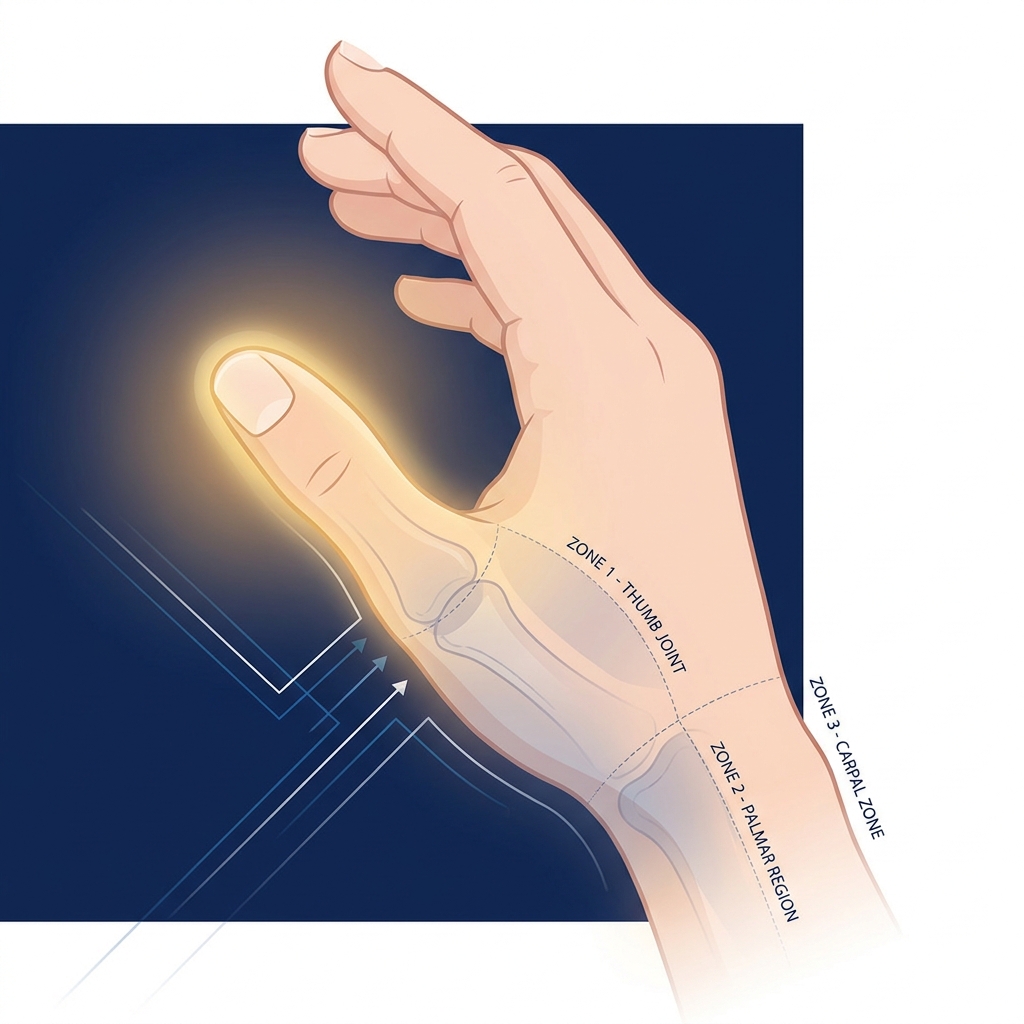
Thumb Pain Joint Treatment: The 3-Condition Diagnosis Map That Matches Your Exact Cause to the Right Regenerative Protocol in 2026
Introduction: Why Your Thumb Pain Keeps Getting Misdiagnosed
Consider a common scenario: a woman in her early fifties is told she has “thumb arthritis.” She receives a cortisone injection, feels better for a few months, and returns for another. By the third visit, the relief lasts weeks instead of months. Something isn’t adding up, and yet no one has questioned whether the original diagnosis was even correct.
This situation plays out constantly, because thumb-side pain is not a single condition. Three distinct diagnoses, basal joint (CMC) arthritis, de Quervain’s tenosynovitis, and trigger thumb, are routinely confused with one another. They share anatomical proximity along the thumb side of the hand and produce overlapping symptoms, which makes misdiagnosis frequent and effective treatment elusive.
The stakes are significant. Loss of thumb function alone imparts a 40 to 50% impairment to the upper extremity because the thumb participates in nearly every grasp and handling task. Getting the right thumb pain joint treatment depends entirely on first identifying which of these three conditions is actually causing the problem.
This article provides a diagnosis-first framework that maps each condition to its own treatment pathway, including where regenerative options fit and why the standard cortisone approach may, in some cases, be quietly making things worse. The goal is to help patients become active, informed participants in their own care decisions.
The Three Conditions Behind Thumb-Side Pain: An Overview
The radial (thumb) side of the wrist and hand is a dense anatomical zone. Joints, tendons, and fibrous sheaths sit in close proximity, which is precisely why symptoms overlap and diagnoses get confused. Three separate conditions dominate this region, each with a completely different underlying problem:
- Basal joint (CMC) arthritis: cartilage degeneration at the joint at the base of the thumb.
- De Quervain’s tenosynovitis: inflammation of the tendon sheath along the first dorsal compartment of the wrist.
- Trigger thumb: constriction of the flexor tendon sheath causing mechanical catching and locking.
These conditions can, and often do, co-occur. A single patient may have CMC arthritis and trigger thumb simultaneously, which compounds the diagnostic puzzle.
The prevalence data underscores why this matters. CMC osteoarthritis affects up to 85% of adults aged 71 to 80 (Journal of Hand Surgery Global Online). De Quervain’s affects roughly 1.3% of women and 0.5% of men in the working population. Trigger thumb carries a lifetime risk of 2.6% and is the fourth most common reason for referral to hand surgeons.
Getting the diagnosis right is the prerequisite for getting the treatment right. A misdiagnosis leads to ineffective interventions and delayed recovery.
Condition 1: Basal Joint (CMC) Arthritis — When the Foundation of the Thumb Wears Down
The carpometacarpal (CMC) joint is the saddle-shaped joint at the base of the thumb, where the thumb metacarpal meets the trapezium bone of the wrist. This joint provides the thumb its remarkable range of motion, but that mobility comes at the cost of stability, making it especially vulnerable to wear.
In CMC arthritis, cartilage progressively degrades until bone contacts bone. Joint instability develops, bone spurs (osteophytes) form, and chronic pain sets in. These are the hallmarks of osteoarthritis. Understanding what makes osteoarthritis worse can help patients make lifestyle adjustments that slow progression.
The condition is extraordinarily common. It affects approximately 15% of adults over age 30, rising to 85% of patients aged 71 to 80. About one-third of postmenopausal women are affected, and two-thirds of women over 55 show radiographic signs. CMC OA is three to four times more prevalent in women than men, with peak impact in postmenopausal women, likely due to hormonal changes affecting ligament laxity.
Clinicians classify severity using the Eaton-Littler staging system (Stages I through IV):
- Stage I: normal or slightly widened joint space.
- Stage II: mild joint space narrowing with small bone spurs.
- Stage III: significant narrowing, large osteophytes, and hardening of bone (sclerosis).
- Stage IV: arthritis has spread to surrounding joints (pantrapezial arthritis).
Importantly, radiographic severity does not always match how a patient feels. Some people with Stage II changes have severe pain, while others with Stage III remain relatively functional. Notably, the CMC joint has been reported as the most painful joint among all hand joints affected by osteoarthritis.
Recognizing CMC Arthritis: Symptom Location and Pattern
- Pain location: deep aching at the base of the thumb, over the thenar eminence (the fleshy mound at the thumb’s base). Patients often describe it as coming from “inside” the joint.
- Aggravating activities: pinching, gripping, turning keys, opening jars, and writing; any task requiring thumb opposition or lateral pinch.
- Characteristic sign: pain and a grinding sensation (crepitus) when the thumb is loaded along its axis, which is the basis of the clinical “grind test.”
- Other features: morning stiffness that eases with movement, visible bony prominence at the thumb base in advanced stages, and weakness in grip and pinch.
How it differs: CMC pain is centered at the joint itself, not along the tendon on the back of the wrist (as in de Quervain’s), and it does not produce catching or locking (as in trigger thumb).
Condition 2: De Quervain’s Tenosynovitis — When the Tendons, Not the Joint, Are the Problem
De Quervain’s tenosynovitis is a stenosing inflammation of the first dorsal compartment of the wrist. It specifically affects the abductor pollicis longus (APL) and extensor pollicis brevis (EPB) tendons as they pass through a fibrous tunnel near the radial styloid.
The pathology is fundamentally a tendon problem, not a joint problem. Repetitive motion causes the tendon sheath to thicken and constrict, creating friction and pain whenever the thumb moves. This type of chronic tendonitis can be particularly stubborn when the underlying mechanical cause is not addressed.
De Quervain’s affects 1.3% of women and 0.5% of men in the working population, with female prevalence six to ten times higher than in men, peaking between ages 40 and 60. A notable modern trend is the rising incidence tied to smartphone and device use, sometimes called “texting tenosynovitis” or “Blackberry Thumb,” which makes the condition increasingly relevant to younger demographics. Other risk factors include new motherhood (repeatedly lifting an infant), musicianship, office work, and racket sports.
The primary clinical diagnostic tool is the Finkelstein test: the patient tucks the thumb inside a closed fist, then the examiner bends the wrist toward the little finger (ulnar deviation). A positive test reproduces sharp pain along the radial wrist.
Recognizing De Quervain’s: Symptom Location and Pattern
- Pain location: along the thumb side of the wrist, at or just above the radial styloid, not at the thumb joint itself.
- Pain character: sharp, burning, or aching pain that may radiate up the forearm or down into the thumb, sometimes with visible swelling over the radial styloid.
- Aggravating activities: pinching, gripping, lifting with the thumb extended, wringing motions, and scrolling on a phone.
- Key distinguishing feature: pain is provoked by moving the thumb away from the hand and by ulnar deviation of the wrist, not by loading the joint.
How it differs: there is no catching or locking (distinguishing it from trigger thumb) and no grinding at the joint base (distinguishing it from CMC arthritis). Tenderness sits directly over the first dorsal compartment.
Condition 3: Trigger Thumb — When the Tendon Gets Stuck
Trigger thumb, or stenosing flexor tenosynovitis, occurs when the flexor pollicis longus tendon becomes constricted within the A1 pulley at the base of the thumb. The result is catching, locking, or a “triggering” sensation during bending and straightening.
The mechanism is mechanical: either the tendon develops a nodule or the pulley thickens, producing a size mismatch that prevents smooth gliding. This is entirely distinct from joint degeneration or dorsal tendon inflammation. Understanding what causes tendon degeneration helps explain why some patients are more susceptible to these mechanical changes.
Trigger thumb carries a lifetime risk of 2.6%, and the thumb is the most frequently affected digit. Risk factors include diabetes (a substantial elevation in risk), rheumatoid arthritis, repetitive gripping, female sex, and age 40 to 60. According to StatPearls, initial management is typically conservative, with rest and splinting for four to six weeks.
Severity ranges from mild catching to a thumb locked in flexion that requires the other hand to straighten it. The Quinnell grading system captures this spectrum, from Grade 0 (no triggering) through Grade 4 (locked and not passively correctable).
Recognizing Trigger Thumb: Symptom Location and Pattern
- Pain location: at the base of the thumb on the palm (volar) side, over the A1 pulley; distinctly different from the dorsal/radial pain of de Quervain’s or the joint pain of CMC arthritis.
- Characteristic symptom: catching, clicking, or locking when bending or straightening the thumb, which is the defining feature.
- Other features: morning stiffness and triggering that eases as the tendon warms, plus a palpable nodule or tenderness at the palm-side base of the thumb.
How it differs: there is no grinding at the joint (distinguishing it from CMC arthritis) and no pain with the Finkelstein test (distinguishing it from de Quervain’s). Ultrasound can confirm the diagnosis by visualizing the thickened pulley and tendon nodule.
A Symptom Map to Identify the Likely Diagnosis
This framework is not a substitute for professional evaluation. It is a tool to help patients communicate more effectively with their provider and understand what questions to ask. A simple three-question pathway narrows things down considerably:
- Where exactly is the pain? Joint base (CMC arthritis) versus radial wrist (de Quervain’s) versus palm side of the thumb base (trigger thumb).
- What makes it worse? Pinching and gripping (CMC) versus wrist deviation and thumb extension (de Quervain’s) versus bending and straightening the thumb (trigger thumb).
- Is there catching or locking? Yes strongly suggests trigger thumb.
| Feature | CMC Arthritis | De Quervain’s | Trigger Thumb |
|---|---|---|---|
| Pain location | Base of thumb, inside joint | Radial wrist, over styloid | Palm side of thumb base |
| Aggravating movement | Pinching, gripping | Thumb extension, wrist deviation | Bending/straightening thumb |
| Characteristic sign | Grinding (crepitus) | Swelling over styloid | Catching/locking |
| Positive test | Grind test | Finkelstein test | Triggering/locking |
| Pain quality | Deep, aching | Sharp, burning | Clicking, mechanical |
Overlap is real. A patient can have two or even all three conditions at once, which is why professional imaging (X-ray for CMC staging, ultrasound for tendon assessment) matters for a definitive diagnosis. Anyone whose symptoms do not fit neatly into one column should seek evaluation from a hand specialist or a regenerative medicine provider who uses diagnostic ultrasound.
The Standard Treatment Ladder and Where It Falls Short
Most patients encounter a predictable treatment progression: rest and activity modification, splinting, NSAIDs, corticosteroid injections, and finally surgery.
Conservative care genuinely helps many patients, particularly in early-stage disease. Cohort studies have shown positive outcomes for nonsurgical treatment at more than five years of follow-up. Yet the surgical escalation reality is sobering: 15 to 30% of CMC OA patients proceed to operative management within one to two years of starting non-operative care, most commonly because pain continues.
The critical limitation lies in corticosteroid injections. They provide temporary relief, typically three to six months, with diminishing returns on repeated use. Most guidelines recommend no more than three to four injections per joint per year. For de Quervain’s and trigger thumb, corticosteroids are also commonly used but carry similar risks of tendon weakening with repeated injection.
The Corticosteroid Trap: Why Repeated Injections May Be Making Things Worse
Corticosteroids reduce inflammation rapidly by suppressing inflammatory cytokines, which is exactly why they deliver quick relief. The problem is what else they do. Corticosteroids also inhibit chondrocyte (cartilage cell) activity, reduce proteoglycan synthesis, and have been shown in laboratory studies to be directly cytotoxic to cartilage cells. In other words, they may suppress the very cellular activity needed to maintain cartilage.
Clinical evidence indicates that repeated intra-articular corticosteroid injections may accelerate cartilage volume loss, particularly beyond three to four injections per year. This concern is significant enough that the Karolinska Institutet’s active randomized trial on PRP for thumb base OA cites steroid-induced cartilage degeneration as a rationale for investigating alternatives.
The trade-off is stark: cortisone may reduce pain today while potentially advancing the disease that causes pain tomorrow. This is not an argument against medicine; it is an evidence-informed reason to explore options that support tissue health rather than suppress it, and to consider them earlier in the treatment journey. For de Quervain’s and trigger thumb, the same logic applies, with the added risks of tendon atrophy and rupture from repeated steroid exposure.
Regenerative Treatment Pathways: Matching the Protocol to the Diagnosis
Regenerative therapies work differently from steroids. Rather than simply suppressing symptoms, they aim to promote tissue repair and reduce inflammation through biological mechanisms. The guiding principle is that the right protocol depends on the specific diagnosis, the stage of disease (for CMC arthritis), and the patient’s individual health profile.
Three modalities feature prominently:
- PRP (Platelet-Rich Plasma): concentrated platelets from the patient’s own blood, rich in growth factors.
- BMAC (Bone Marrow Aspiration Concentrate): concentrated bone marrow cells containing mesenchymal stem cells and anti-inflammatory factors.
- Exosome therapy: extracellular vesicles that carry signaling molecules between cells.
Every regenerative injection for a thumb condition should be delivered under ultrasound guidance to ensure it reaches the correct anatomical target, whether the CMC joint, the tendon sheath, or the A1 pulley. This is especially important given the small, complex anatomy of the thumb. Unicorn Bioscience uses precision imaging guidance (ultrasound and X-ray) for all injection procedures, a meaningful differentiator for small joint and tendon treatments.
Regarding regulation: as of 2026, the FDA has not approved PRP, stem cell, or exosome products specifically for orthopedic conditions, but substantial clinical evidence supports safety and efficacy when administered by qualified providers within FDA regulatory frameworks.
Regenerative Protocol for Basal Joint (CMC) Arthritis
Eaton staging guides treatment selection. Regenerative therapies are most appropriate and most effective for Eaton Stages I and II, where cartilage remains and the biological environment can support repair. Stage III may still benefit with realistic expectations, while Stage IV typically warrants surgical consultation.
The evidence is encouraging. A Mayo Clinic tertiary care study found that 68.8% of injected joints reported moderate or excellent symptom improvement, with a mean benefit duration of 15.6 months (PMC, Mayo Clinic). PRP delivers concentrated growth factors (PDGF, TGF-beta, VEGF) that stimulate chondrocyte activity and reduce inflammatory cytokines. A 2023 systematic review found that PRP and lipofilling show promising results for basal thumb arthritis, and a 2025 systematic review and meta-analysis confirmed PRP as a promising alternative with encouraging outcomes.
BMAC contains mesenchymal stem cells, growth factors, and anti-inflammatory cytokines, offering a more potent regenerative signal for more advanced cartilage loss. Understanding the differences between BMAC and stem cell therapy can help patients and providers select the most appropriate option. Adipose-derived stem cell therapy (liparthroplasty) is another option: a 31-patient case series showed median VAS pain scores fall from 7 to 2 at two years, with lipoaspirate providing both regenerative and cushioning effects. Exosome therapy modulates inflammation and promotes regeneration, and may suit patients who are not ideal candidates for autologous cell therapies.
This is the “regenerative bridge” concept: PRP and cell-based therapies may delay or eliminate the need for surgery, particularly for Eaton Stage I and II patients who are failing conservative care but are not yet surgical candidates.
Regenerative Protocol for De Quervain’s Tenosynovitis
De Quervain’s is a tendon sheath condition, so the regenerative target is the first dorsal compartment sheath, not the joint. Standard first-line care remains appropriate: thumb spica splinting, activity modification, NSAIDs, and physical therapy. Corticosteroid injection is common but risks tendon weakening with repeated use.
PRP injected into the tendon sheath delivers growth factors that promote tenocyte proliferation and sheath remodeling, addressing the underlying pathology rather than masking inflammation. Ultrasound guidance is essential here because the first dorsal compartment often has a septum separating the APL and EPB sub-compartments, and missing it is a common cause of treatment failure. This approach is particularly compelling for patients who have already had one or two steroid injections with diminishing relief. Activity modification, including changes in smartphone habits, is an essential complement to any injection therapy.
Regenerative Protocol for Trigger Thumb
Trigger thumb involves the A1 pulley and flexor tendon sheath on the palm side of the thumb, a completely different anatomical target. The standard treatment ladder runs from splinting and rest (four to six weeks) to corticosteroid injection to percutaneous or open pulley release surgery. Corticosteroid injection works well in many Quinnell Grade I and II cases, but success declines with repeated injections and in patients with diabetes.
PRP may reduce sheath inflammation and promote remodeling of the thickened A1 pulley without the tissue-weakening risks of steroids. Exosome injection for tendon healing represents another emerging option that may support tissue repair at the cellular signaling level. Ultrasound-guided injection is essential for accuracy, and ultrasound doubles as a diagnostic tool by visualizing pulley thickening and tendon nodules. The evidence base for regenerative therapy in trigger thumb is less mature than for CMC arthritis, but the biological rationale and safety profile make it a reasonable option for patients who have failed or are poor candidates for corticosteroid injection. For severe or locked trigger thumb (Quinnell Grade III and IV), surgical consultation may be appropriate.
The Role of Ultrasound-Guided Precision in Thumb Regenerative Injections
Image guidance is not optional for thumb regenerative injections. The CMC joint, first dorsal compartment, and A1 pulley are small, anatomically complex structures where blind injection accuracy falls well short of ultrasound-guided placement.
The clinical significance is direct: if the regenerative agent misses the target tissue, therapeutic benefit is reduced or lost entirely. That is a particular concern with biologics like BMAC or exosomes. Ultrasound provides real-time visualization of the needle tip, the target structure, and the surrounding anatomy, allowing the provider to confirm accurate placement before injecting. For CMC joint injections, X-ray (fluoroscopy) may confirm intra-articular placement, especially when joint space is significantly narrowed.
Unicorn Bioscience’s use of both ultrasound and X-ray guidance is a key clinical differentiator that directly affects outcomes. Ultrasound also plays a diagnostic role, identifying tendon sheath thickening (de Quervain’s), pulley thickening and nodules (trigger thumb), and joint effusion or synovitis (CMC arthritis).
Building a Personalized Thumb Pain Treatment Plan: What to Expect
A comprehensive evaluation at a regenerative medicine clinic generally includes a history and symptom review, a physical examination (grind test, Finkelstein test, triggering assessment), an imaging review (X-ray for Eaton staging, ultrasound for tendon assessment), and a discussion of treatment goals.
Personalized protocols are built around multiple factors: diagnosis and disease stage, inflammation levels, patient age, overall health, current medications (including anticoagulants that may affect PRP processing), and activity goals. Some patients benefit from combination therapy, such as PRP for the CMC joint paired with a hyaluronic acid injection for viscosupplementation, or PRP alongside physical therapy for de Quervain’s.
Realistic expectations matter. Regenerative therapies are not instant fixes. Most patients see progressive improvement over 4 to 12 weeks as biological repair unfolds, and some require a series of injections. Same-day treatment is available for qualified candidates at Unicorn Bioscience, and virtual consultations allow patients to discuss their case before committing to an in-person visit, which is convenient for those near any of the clinic’s eight locations.
When Regenerative Therapy Is Most Likely to Help and When Surgery May Still Be Necessary
Regenerative care is not a universal solution. It works best when matched to the right patient at the right stage.
- CMC arthritis: most appropriate for Eaton Stage I and II (and selected Stage III) patients who have failed conservative care but are not yet at the point where surgery is the only option. Stage IV with complete joint destruction typically requires surgical consultation.
- De Quervain’s: best suited to patients who have failed splinting and one or two steroid injections, or who want to avoid repeated steroid exposure. Surgical release is effective for refractory cases.
- Trigger thumb: most appropriate for Quinnell Grade I and II. Grade III and IV (locked thumb) may require surgical pulley release.
The goal is not to replace surgery in every case but to provide an effective intermediate option that may delay or eliminate the need for it. Reflecting the potential of this approach, more than 90% of Unicorn Bioscience’s stem cell patients have not gone on to joint replacement surgery. Patients should seek a provider willing to give an honest assessment, because not everyone is a candidate, and a reputable clinic will say so.
Frequently Asked Questions About Thumb Pain Joint Treatment
How do I know if my thumb pain is arthritis or a tendon problem?
Consider pain location (joint base versus radial wrist versus palm side), the movements that aggravate it, and whether there is catching or locking. Joint-base grinding suggests arthritis; radial-wrist pain with a positive Finkelstein test suggests de Quervain’s; catching or locking points to trigger thumb.
Can a patient have more than one of these conditions at the same time?
Yes. CMC arthritis, de Quervain’s, and trigger thumb can coexist, which is exactly why a thorough evaluation including ultrasound is important.
How many cortisone injections are too many?
Most guidelines recommend no more than three to four per joint per year. Beyond that, the risk of cartilage damage and tendon weakening rises significantly.
Is PRP safe for the thumb?
Mayo Clinic data indicate PRP is a relatively safe treatment for thumb CMC arthritis, and because it uses the patient’s own blood, immunological risk is minimal.
How long does PRP relief last for thumb arthritis?
The Mayo Clinic study reported a mean benefit duration of 15.6 months, though individual results vary with disease stage and patient factors.
Do patients with Stage III CMC arthritis need surgery?
Not necessarily. Many Stage III patients benefit from regenerative therapy. Surgical consultation is appropriate when conservative and regenerative care have failed.
What is the Eaton staging system and why does it matter?
It classifies CMC arthritis into four progressive radiographic stages. Staging guides treatment because earlier stages are better candidates for regenerative therapy.
Are regenerative treatments FDA-approved for thumb conditions?
As of 2026, the FDA has not approved PRP, stem cells, or exosomes specifically for orthopedic conditions, but substantial clinical evidence supports safety and efficacy when administered by qualified providers within FDA regulatory frameworks.
Conclusion: The Right Diagnosis Is the First Step to the Right Treatment
Thumb-side pain is not a single condition, and treating it as one leads to mismatched interventions and prolonged suffering. The three-condition framework brings clarity: basal joint arthritis (joint degeneration), de Quervain’s tenosynovitis (dorsal tendon sheath inflammation), and trigger thumb (volar flexor tendon constriction) each present with distinct symptom patterns, diagnostic tests, and treatment pathways.
The corticosteroid trap deserves particular attention. Repeated cortisone injections may provide short-term relief while potentially accelerating long-term cartilage damage, a compelling reason to consider regenerative alternatives earlier. The evidence is building: PRP for CMC arthritis shows 68.8% moderate-to-excellent improvement with 15.6 months of mean benefit, adipose-derived stem cell therapy shows significant pain reduction at two years, and a 2024 Karolinska randomized trial is actively strengthening the evidence base.
The best outcomes come from precision: the right diagnosis, the right regenerative protocol, delivered to the correct anatomical target under image guidance. Patients who understand their diagnosis and their options are far better equipped to make informed decisions and to advocate for thumb pain joint treatment that addresses the root cause rather than just the symptoms. For those also dealing with related joint issues in the wrist, exploring wrist arthritis non-surgical treatment options may provide additional context for a comprehensive hand and wrist care plan.
Take the Next Step: Get a Personalized Thumb Pain Evaluation at Unicorn Bioscience
Patients experiencing persistent thumb pain are invited to schedule a consultation with Unicorn Bioscience for a diagnosis-first evaluation and a personalized treatment plan. Both virtual and in-person consultations are available, making expert assessment accessible regardless of location.
Unicorn Bioscience operates eight locations across Texas (Austin, Dallas, El Paso, Fort Worth, Houston, and San Antonio), Florida (Boca Raton), and New York (Manhattan). Key differentiators relevant to thumb pain include precision ultrasound and X-ray guided injections, multi-modal regenerative options (PRP, BMAC, and exosome therapy), personalized protocols based on individual patient factors, and same-day treatment availability for qualified candidates. The team includes providers with training from prestigious institutions including Johns Hopkins.
To learn more, call (737) 347-0446 or visit unicornbioscience.com.
Schedule Your Consultation Today!
