
What Causes Tendon Degeneration: The 5-Factor Biological Cascade That Explains Why Tendons Fail to Heal
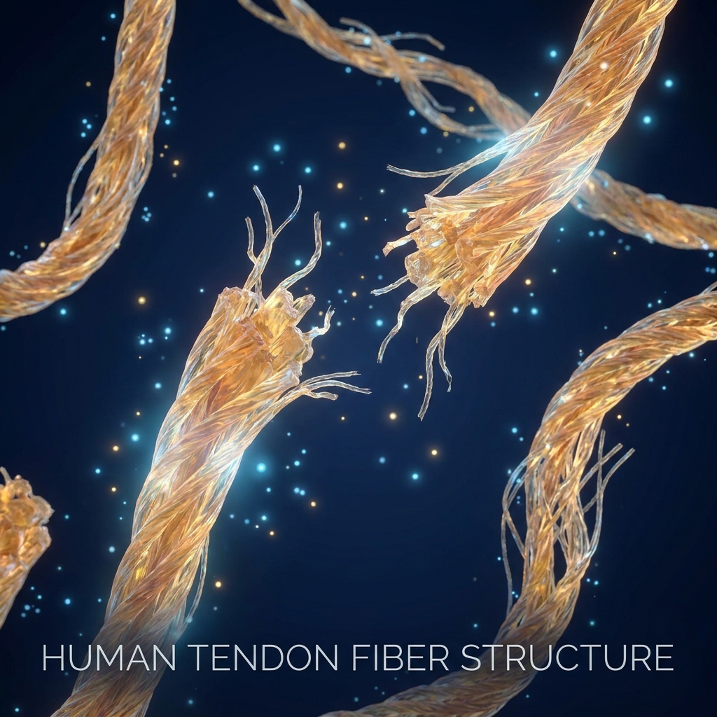
What Causes Tendon Degeneration: The 5-Factor Biological Cascade That Explains Why Tendons Fail to Heal
Introduction: Why ‘Overuse’ Is an Incomplete Answer
A patient receives a diagnosis of tendinopathy and is told simply to rest and avoid overuse. Months later, the pain returns, often worse than before. This frustrating cycle plays out in clinics across the country every day, leaving patients confused about why their tendons refuse to heal.
The core problem lies in how tendon degeneration is typically explained. Conventional patient education reduces this complex condition to a single cause, leaving patients without the understanding needed to make informed treatment decisions. The reality is far more nuanced: tendon degeneration is not a single-cause event but a multifactorial biological cascade involving mechanical overloading, failed healing biology, oxidative stress, metabolic amplifiers, and drug-induced damage.
Before exploring this cascade, clarifying terminology is essential. Tendinitis refers to an inflammatory response, while tendinosis describes degenerative changes with little to no inflammation. Tendinopathy serves as the umbrella term encompassing both conditions, which often coexist along a continuum of pathology. Most patients still believe all tendon pain is purely inflammatory, which explains why anti-inflammatory drugs so often fail to provide lasting relief.
This article offers a different approach. Each biological root cause will be connected to why regenerative therapies such as PRP, stem cells, and exosomes address actual failure points rather than simply masking pain. The scope of this problem is substantial: approximately 30% of all musculoskeletal consultations in general practice are tendinopathy-related, and 30 to 50% of all sporting injuries involve tendon pathology. Yet up to two-thirds of cases go unreported, meaning the true burden is likely much higher than current statistics suggest.
Understanding the Tendon: Structure, Function, and Inherent Vulnerability
Tendons are remarkable structures composed of roughly 65 to 80% Type I collagen organized in a tightly hierarchical matrix. Tenocytes and tenoblasts comprise 90 to 95% of cellular elements, and this precise organization gives tendons their exceptional tensile strength.
Tenocytes serve as the primary cells responsible for synthesizing, maintaining, and remodeling the extracellular matrix (ECM). Their health is central to every stage of the degeneration cascade. When tenocytes function properly, they continuously repair and maintain the collagen architecture that allows tendons to withstand tremendous mechanical forces.
However, tendons possess an inherent vulnerability: natural hypovascularity. Compared to most tissues, tendons have a poor intrinsic blood supply, making them inherently slow to receive nutrients, oxygen, and healing signals. This is not a pathological finding but a baseline vulnerability that sets the stage for problems when injury occurs.
Because tendons are structurally dependent on tenocyte health and collagen organization, and because they are naturally under-resourced in terms of blood supply, any disruption at the cellular or vascular level has outsized consequences. Understanding this vulnerability is critical for grasping why tendinopathy progresses through three stages: reactive tendinopathy (still reversible), tendon disrepair (accumulating damage), and degenerative tendinopathy (involving irreversible cell death, matrix disorganization, and high rupture risk).
Factor 1: Mechanical Overloading and the Tenocyte-Mediated Breakdown Cascade
The primary mechanical trigger for tendon degeneration is excessive loading that surpasses the tendon’s adaptive capacity. This is commonly summarized as “too much, too soon,” leading to microruptures in the collagen matrix.
At the cellular level, mechanical overloading activates stressed tenocytes to release pro-inflammatory cytokines including IL-1β, TNF-α, and PGE2, initiating a local inflammatory response. This cytokine signaling upregulates matrix metalloproteinases (MMP-1, MMP-13) that actively degrade Type I collagen while simultaneously inhibiting new collagen synthesis.
This process is not simply “wear and tear.” It is an active, cell-mediated breakdown process where the tendon’s own cellular machinery turns against its structural integrity. According to research published in the Journal of Musculoskeletal Surgery and Research, excessive mechanical loading that surpasses the tendon’s adaptive capacity is the primary contributing factor to tendinopathy.
A common misconception is that tendinopathy only affects athletes. While athletes face high-load overuse, sedentary workers and manual laborers develop tendinopathy through repetitive low-load stress that accumulates over time. The cascade operates identically regardless of activity level, and inflammation and degeneration are not mutually exclusive but work together in tendinopathy pathogenesis. Prevalence data shows tendinopathy is nearly twice as common in athletes with extremely high training exposure. Achilles and patellar tendinopathies are the most common lower-limb presentations (6.2% each), while rotator cuff tendinopathy leads upper-limb cases at 6.02%.
Regenerative therapies such as PRP and stem cell therapy address this failure point by delivering growth factors (PDGF, IGF, VEGF) that restore healthy tenocyte signaling and support matrix repair, directly countering the MMP-driven breakdown cycle.
Factor 2: Poor Vascularity and the Failed Healing Loop
When a tendon sustains microruptures, its limited blood supply means healing signals, immune cells, and nutrients arrive slowly and in insufficient quantities. The consequence of this failed healing response is significant: instead of regenerating healthy Type I collagen, the tendon produces inferior Type III scar collagen that is structurally weaker, less organized, and more prone to re-injury.
A paradox emerges in chronic tendinopathy. The body attempts to compensate by growing new blood vessels (neovascularization) into normally avascular tendon regions. However, this abnormal vasculature is accompanied by neural in-growth, which is the primary mechanism behind chronic tendon pain that persists even after inflammation has resolved. Research from Advanced Science explores the complex interplay between VEGF, HIF-1, and microRNA regulation of angiogenesis in driving this abnormal process.
This creates a self-reinforcing loop: poor healing leads to scar collagen accumulation, which causes structural weakness, which results in re-injury, which perpetuates continued failed healing. Exosome therapy and PRP deliver VEGF and other angiogenic growth factors that support healthy vascular remodeling rather than the chaotic neovascularization seen in chronic tendinopathy. Stem cells modulate the vascular microenvironment through paracrine signaling.
Factor 3: Reactive Oxygen Species and Tenocyte Apoptosis
Reactive oxygen species (ROS) require nuanced understanding. At physiological levels, ROS support cellular signaling and are a normal part of tendon biology. The problem arises when ROS accumulate beyond the tendon’s antioxidant defense capacity.
Multiple sources drive excess ROS in tendinopathy: mechanical overload, hypoxia from poor vascularity, metabolic disease, and aging all independently contribute. This means multiple factors in this cascade converge on the same oxidative damage pathway. Research confirms that ROS play a dual role, regulating cellular processes under physiological conditions while contributing to tendinopathy and impaired healing when dysregulated.
The downstream consequences of excess ROS are severe. They trigger tenocyte apoptosis (programmed cell death), degrade extracellular matrix components, and impair the tendon’s ability to mount an effective healing response. Recent research has identified a newly significant mechanism: mechanical overuse may hyperactivate calcium channels, leading to chronically elevated intracellular calcium that further amplifies ROS production. This creates a cycle that accelerates degeneration independent of continued mechanical stress.
Conventional NSAIDs and corticosteroids do not address oxidative stress or tenocyte apoptosis. Regenerative therapies, particularly exosomes and MSC-based treatments, modulate the oxidative microenvironment and deliver anti-apoptotic signaling molecules that help restore tenocyte viability.
Factor 4: Metabolic Disorders — How Diabetes, Obesity, and Dyslipidemia Amplify Every Step
Metabolic disorders do not cause tendinopathy in isolation but dramatically accelerate and worsen every biological failure point already described.
Diabetes presents a particularly significant risk. A 2025 meta-analysis published in PMC/NIH showed diabetes raises the odds of Achilles tendinopathy by an odds ratio of 7.22. The mechanism involves hyperglycemia altering collagen cross-linking and structure, impairing ischemic tolerance, and compounding oxidative stress through advanced glycation end-products (AGEs).
Obesity is present in approximately 64% of tendinopathy patients. The dual mechanism involves increased mechanical load on tendons combined with systemic low-grade inflammation driven by adipose tissue cytokines (adipokines).
Dyslipidemia and hypercholesterolemia cause excess lipid deposition that creates collagen fibril disorganization within the tendon matrix, directly disrupting the structural architecture that gives tendons their tensile strength.
Smoking reduces oxygen delivery to tendons, compounding the existing hypovascularity problem and worsening the failed healing loop.
For older patients, the concept of “inflammaging” becomes relevant. Aging tendons exhibit mitochondrial dysfunction, decreased mechanical strength, increased glycosaminoglycans, slower healing rates, and a chronic low-grade inflammatory state. This makes metabolic comorbidities particularly dangerous in patients over 40.
This section addresses an underrepresented patient population: tendinopathy is not only a sports injury. Sedentary and metabolically compromised patients face significant risk through entirely non-athletic mechanisms. MSC efficacy is reduced by aging and metabolic disease, which is why personalized treatment protocols that account for patient age, inflammation levels, and metabolic status are essential for optimizing outcomes.
Factor 5: Drug-Induced Tendon Degeneration — The Risk Most Patients Are Never Told About
Drug-induced tendinopathy is a clinically significant, FDA-recognized cause of tendon degeneration that is almost entirely absent from patient-facing content.
Fluoroquinolone antibiotics (ciprofloxacin, levofloxacin) carry FDA black-box warnings for tendon rupture risk. Research explains that these commonly prescribed antibiotics decrease collagen and proteoglycan synthesis, disrupt tendon regeneration through downregulation of MMP-mediated remodeling, and cause direct tenocyte toxicity.
Corticosteroids, while frequently injected for tendon pain, decrease tenocyte viability, alter matrix synthesis, and promote apoptosis. Repeated corticosteroid injections may provide short-term pain relief while actively accelerating the underlying degeneration.
The clinical irony is notable: two of the most commonly prescribed treatments for tendon pain (corticosteroid injections) and infections in active patients (fluoroquinolones) are independently capable of worsening the biological cascade they are meant to address.
Statins represent an emerging concern. Dyslipidemia treatment with statins has also been associated with tendon pathology, though the evidence is less definitive than for fluoroquinolones and corticosteroids.
Patients should not discontinue prescribed medications without medical guidance. However, they should be informed of these risks so they can have informed conversations with their providers and understand why their tendon condition may not be improving. Because corticosteroids and fluoroquinolones damage the very cellular machinery that tendons need to heal, regenerative therapies that restore tenocyte health, promote collagen synthesis, and modulate the ECM represent a logical therapeutic response to drug-induced degeneration.
Why Conventional Treatments Fall Short: The Biology of Incomplete Recovery
NSAIDs address pain and acute inflammation but do not repair structural damage, restore Type I collagen architecture, reverse tenocyte apoptosis, or address oxidative stress. They are appropriate for symptom management but insufficient as a standalone treatment for degenerative tendinopathy.
Repeated corticosteroid injections may reduce pain short-term but accelerate the biological cascade, explaining the common clinical pattern of temporary relief followed by worsening symptoms. Research from The Journal of Physiology confirms that NSAIDs and corticosteroids do not contribute to long-term recovery.
Rest alone is insufficient for advanced tendinopathy. Once the tendon has progressed to the disrepair or degenerative stage, passive rest does not reverse the accumulated cellular death, scar collagen deposition, or matrix disorganization. Because tendons rely on tenocyte activity and growth factor signaling to regenerate, and because these systems are compromised in degenerative tendinopathy, the tendon cannot self-repair past a certain threshold without biological intervention.
How Regenerative Therapies Address the Biological Root Causes
Regenerative therapies work not by masking pain but by targeting the specific biological failure points identified throughout this article.
PRP (Platelet-Rich Plasma): Restoring Growth Factor Signaling
PRP concentrates the patient’s own platelets, which upon activation release growth factors including PDGF, IGF, VEGF, and TGF-β. These are the same signaling molecules that tenocytes lose access to during degeneration.
PRP directly addresses Factor 1 by stimulating tenocyte proliferation and healthy collagen synthesis. It addresses Factor 2 by delivering VEGF to support healthy vascular remodeling. It also modulates the inflammatory microenvironment to reduce the cytokine burden that drives further breakdown. When combined with extracorporeal shockwave therapy (ESWT), PRP enhances growth factor activity at the injury site, facilitating tendon regeneration.
PRP efficacy depends on platelet concentration, patient health status, and delivery precision, which is why imaging-guided injection using ultrasound or X-ray is essential for accurate therapeutic delivery.
Stem Cell Therapy (MSCs): Regenerating the Cellular Foundation
Mesenchymal stem cells (MSCs) from bone marrow (BMAC) or adipose tissue promote tendon regeneration through three pathways: paracrine signaling (releasing healing factors into the local environment), immune modulation (reducing the chronic inflammatory state), and direct differentiation into tendon-lineage cells.
MSCs directly address Factor 3 by delivering anti-apoptotic signals and antioxidant factors. They address Factor 2 by promoting Type I collagen synthesis over inferior Type III scar collagen. They also address the metabolic amplification of Factor 4 by modulating the local inflammatory microenvironment.
Aging reduces MSC efficacy, which is why patient age and metabolic status are critical variables in treatment planning. As of 2026, the FDA has not approved stem cell products specifically for orthopedic conditions, but substantial clinical evidence supports safety and efficacy when administered by qualified providers within FDA regulatory frameworks.
Exosome Therapy: Cellular Communication at the Molecular Level
Exosomes are extracellular vesicles that carry microRNAs, proteins, and signaling molecules between cells. In the context of tendinopathy, they deliver regenerative instructions directly to damaged tenocytes and the surrounding ECM.
Exosomes address the microRNA regulation of angiogenesis (relevant to Factor 2’s neovascularization problem), deliver anti-inflammatory signals that counter the cytokine cascade from Factor 1, and support tenocyte survival against the oxidative stress driving Factor 3. Because they operate at the molecular signaling level, they can modulate the tendon’s biological environment without introducing live cells, potentially offering advantages in patients where MSC efficacy is reduced by age or metabolic disease.
The Importance of Personalized, Precision-Guided Treatment
Because tendon degeneration is multifactorial, no single treatment addresses every failure point. The most effective approach combines therapies matched to the patient’s specific biological profile.
Patient factors that matter for treatment selection include inflammation levels, patient age, injury stage (reactive vs. disrepair vs. degenerative), current medications (especially fluoroquinolones or corticosteroids), metabolic health (diabetes, obesity, dyslipidemia), and personal health goals. Accurate injection placement using ultrasound or X-ray guidance is not a luxury but a clinical necessity. Regenerative therapies can only address biological failure points if they are delivered precisely to the affected tissue.
Early intervention is imperative. Because the three-stage model shows that early-stage (reactive) tendinopathy is still reversible while late-stage degeneration involves irreversible cell death, understanding the biology of degeneration provides the rationale for seeking evaluation before symptoms become chronic.
The socioeconomic dimension is also significant. Shoulder tendinopathy takes approximately 10 months to heal on average, and about a quarter of tennis elbow patients report difficulty with daily activities including dressing, carrying objects, and driving. The burden of undertreated tendinopathy extends well beyond athletic performance. For patients dealing with tennis elbow specifically, PRP therapy for tennis elbow has demonstrated meaningful success rates in clinical settings.
Conclusion: From Root Cause to Real Recovery
The five-factor biological cascade can be summarized as follows: mechanical overloading triggers tenocyte-mediated MMP upregulation and collagen breakdown; poor vascularity traps the tendon in a failed healing loop producing inferior scar collagen; reactive oxygen species accumulate and drive tenocyte apoptosis; metabolic disorders amplify every step; and certain commonly prescribed drugs actively accelerate degeneration.
Understanding what causes tendon degeneration at a biological level is not purely educational. It is the foundation for making treatment decisions that address root causes rather than temporarily masking symptoms. The conventional approach (pain management without structural repair) contrasts sharply with the regenerative approach (targeting specific biological failure points with PRP, stem cells, and exosomes).
Regenerative medicine is not a guaranteed cure, and outcomes depend on injury stage, patient health, and treatment precision. However, the mechanistic rationale for these therapies is grounded in the same biology that explains why conventional treatments fall short. Patients who understand the biology of their condition are better equipped to ask the right questions, evaluate their treatment options, and advocate for care that goes beyond symptom suppression.
Ready to Address the Root Cause of Tendon Pain?
For patients whose experience resonates with the biological cascade described in this article, including chronic pain that returns after rest, incomplete recovery from conventional treatment, or known metabolic risk factors, a personalized evaluation may reveal whether regenerative therapy is appropriate for their specific situation.
Unicorn Bioscience offers precision-guided regenerative therapies, including PRP, stem cell therapy, BMAC, and exosome therapy, delivered by a team that includes physicians and physician assistants with training from institutions including Johns Hopkins. With locations across Texas, Florida, and New York, the practice provides accessible care for patients seeking alternatives to surgery.
Treatment plans are developed based on individual patient factors including inflammation levels, age, injury type, current medications, and health goals, directly addressing the multifactorial nature of tendon degeneration described throughout this article. Same-day treatment availability and virtual consultation options reduce barriers to the first step.
To discuss a tendon condition and explore whether regenerative medicine is appropriate, schedule a consultation (virtual or in-person) by calling (737) 347-0446 or visiting unicornbioscience.com.
As of 2026, the FDA has not approved stem cell, PRP, or exosome products specifically for orthopedic conditions, but substantial clinical evidence supports safety and efficacy when administered by qualified providers within FDA regulatory frameworks.
Schedule Your Consultation Today!
