
How Is PRP Prepared from a Blood Sample: The Two-Spin Science Framework That Explains Every Step from Vial to Injection in 2026
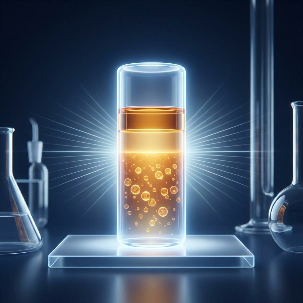
How Is PRP Prepared from a Blood Sample: The Two-Spin Science Framework That Explains Every Step from Vial to Injection in 2026
Introduction: Why “We Spin Your Blood” Is Not Enough of an Answer
A patient sits in a clinic chair, watches a small vial of their own blood disappear into a centrifuge, and roughly twenty minutes later that vial returns transformed. The dark red liquid has become something lighter, clearer, and biologically different. And yet, for most patients, no one explains why.
The typical explanation of platelet-rich plasma (PRP) preparation reduces the process to a tidy three-step summary: draw blood, spin it, inject it. For a scientifically curious patient, that summary raises more questions than it answers. Why does spinning concentrate platelets? What are the layers that form inside the tube? Why do some clinics spin once and others spin twice?
This article offers a physics-first, patient-friendly walkthrough of every step, from the moment blood enters the collection tube to the moment growth factors are released at the injection site. It draws on real centrifugation parameters, cellular biology, and preparation science.
At its foundation, PRP is an autologous blood-derived concentrate, meaning it is built entirely from the patient’s own biology. Through controlled centrifugation, it achieves platelet concentrations 2 to 5 times higher than whole blood. The entire process can be understood through what this article calls the two-spin framework: a logical scientific architecture that governs how a vial becomes a therapy. As of 2026, peer-reviewed research continues to refine and standardize these protocols, making PRP an actively evolving science worth understanding in detail.
What PRP Actually Is: Platelets, Plasma, and the Biology of Healing
PRP is precisely defined as a concentration of platelets suspended in a small volume of plasma, separated from the other cellular components of whole blood. That definition sounds simple, but each word carries biological weight.
Platelets are tiny, anucleate cell fragments (they contain no nucleus) produced by megakaryocytes in the bone marrow. Their primary biological role is hemostasis (the stopping of bleeding) and tissue repair. When tissue is damaged, platelets are among the first responders.
The therapeutic power of PRP originates inside these platelets. Each platelet contains 50 to 80 alpha-granules, and collectively those granules store over 400 bioactive proteins. Among the most important are the growth factors: PDGF (platelet-derived growth factor), TGF-β1, VEGF, bFGF, EGF, IGF-1, and HGF. These proteins orchestrate tissue regeneration, stimulate new blood vessel formation, and recruit repair cells to injured areas. According to a 2025 peer-reviewed review, these molecules are the biological engine behind PRP’s clinical effects.
Concentration is the entire point. According to the Red Cross, PRP is defined as having at least 200,000 platelets per microliter (μL), but clinical PRP targets 1 to 1.5 million platelets/μL for optimal therapeutic effect. At physiological baseline, platelets are present in the blood but not concentrated enough to overwhelm a chronic injury environment. PRP changes that ratio dramatically.
Because the patient’s own baseline platelet count directly affects the final PRP yield, the treatment is inherently personalized. Two patients undergoing the identical protocol can produce different final concentrations based on their individual biology.
Step 1: The Blood Draw: Volume, Timing, and Why the Vial Is Already Doing Something
The process begins with a standard venous blood draw. Typically 10 to 60 mL of peripheral blood is collected, with the exact volume depending on the clinical indication and the preparation system used. A 30 mL draw commonly yields 3 to 5 mL of final PRP product, which helps explain why a relatively large draw is needed to produce a small therapeutic volume.
One detail most patients never hear: the collection tube is not empty. It is pre-loaded with an anticoagulant, and the moment blood enters the tube, that anticoagulant begins working.
Anticoagulation is essential. Without it, platelets would immediately activate and clump together inside the tube, releasing their growth factors prematurely and exactly where those growth factors are not wanted. The goal is to keep the platelets calm and intact until they reach the injection site.
Timing matters as well. Blood samples should be processed promptly after collection to preserve platelet viability and prevent spontaneous activation. Prepared PRP should also be used within a defined window to maintain therapeutic potency, which is precisely why in-clinic preparation and same-day injection is the standard approach at practices like Unicorn Bioscience.
The Anticoagulant Decision: ACD-A, Sodium Citrate, and Why This Chemistry Matters
Most anticoagulants used in PRP work through a shared mechanism: they chelate (or bind) ionized calcium ions. Calcium is an essential cofactor in the coagulation cascade. Remove the calcium, and clotting simply cannot proceed.
ACD-A (Acid Citrate Dextrose Solution A) is the only FDA-approved anticoagulant for PRP preparation, making it the most commonly used option in clinical settings. It preserves platelet morphology well, but there is a trade-off: ACD-A lowers blood pH to approximately 6.5, which can affect the half-life and stability of certain growth factors. This is a nuance patients almost never hear discussed.
Sodium citrate serves as an alternative. Some studies associate it with greater induction of mesenchymal cell proliferation, making it relevant for specific regenerative applications.
EDTA, by contrast, yields the highest raw platelet count but causes platelet swelling and premature activation. Research comparing anticoagulants in PRP preparation found EDTA produced higher platelet recovery but at the cost of platelet integrity, making it unsuitable for therapeutic PRP despite its routine use in diagnostic blood panels.
Anticoagulant selection is therefore a clinical decision, not a default setting. This single variable can meaningfully affect the biological quality of the final product, and patients are entitled to ask which anticoagulant is used and why.
The Physics of Separation: How Centrifugation Works at the Cellular Level
At the heart of PRP preparation lies differential centrifugation. The centrifuge applies controlled acceleration force to exploit differences in the specific gravity (or density) of the various blood components.
In patient-friendly terms: red blood cells are denser and heavier, so they sink faster under force. Platelets are lighter and smaller, so they resist sedimentation at lower forces. By calibrating that force, a technician can selectively move some components while leaving others suspended.
A critical distinction separates informed protocols from imprecise ones: RPM versus g-force. RPM (revolutions per minute) measures how fast the rotor spins, but g-force (also called RCF, or relative centrifugal force) is the scientifically meaningful parameter because it accounts for rotor radius. Two centrifuges spinning at the same RPM can produce entirely different g-forces depending on rotor size. This is why validated protocols always specify g-force, not just RPM.
The density hierarchy of blood components, from heaviest to lightest, runs as follows: red blood cells, granulocytes, monocytes, lymphocytes (which together form the buffy coat), platelets, and finally plasma proteins. By adjusting g-force and duration, a technician can selectively sediment specific layers while leaving others in suspension. As research on centrifugation parameters for PRP confirms, acceleration, time, and processed volume are all critical variables for reproducible results.
A helpful analogy: like a salad dressing that separates into oil and vinegar layers when left undisturbed, centrifugation accelerates this natural density-based separation by thousands of times.
The Two-Spin Framework: A Step-by-Step Walk Through the Double Centrifugation Method
The double-spin (or two-spin) method is widely regarded as the gold-standard preparation approach for achieving high platelet concentration. Two spins outperform one because they divide the work into two distinct, complementary jobs. The first spin separates whole blood into its major layers. The second spin concentrates the platelets drawn from those layers.
The First Spin: Soft Centrifugation and the Three-Layer Separation
The first spin is a “soft spin,” using a lower centrifugal force (typically 100 to 300×g) applied for 10 to 15 minutes. This gentle force is calibrated to sediment red blood cells without pelleting the platelets.
If a patient could watch their tube after this first spin, they would see three distinct layers emerge:
- The bottom red layer: densely packed red blood cells (erythrocytes).
- A thin whitish-gray intermediate band, the buffy coat: rich in white blood cells (leukocytes) and some platelets.
- The upper yellow layer: plasma containing platelets, growth factors, and proteins.
Calibration is everything here. Too much force and platelets begin to pellet with the red blood cells, reducing yield. Too little force and red blood cells remain in suspension, contaminating the platelet layer. A validated high-yield double-spin protocol uses 100×g for 15 minutes as the first spin, part of a protocol achieving 86 to 99% platelet recovery.
The buffy coat is more than a curiosity. This thin intermediate layer serves as the anatomical landmark that guides the next step, marking the boundary between the platelet-rich plasma above and the cellular components below.
Between the Spins: Extracting the Platelet-Rich Layer
Once the first spin is complete, the technician carefully removes the tube from the centrifuge without disturbing the layers. Using a syringe or pipette, the upper plasma layer containing the platelets is carefully drawn off, stopping just above the buffy coat to avoid contaminating the PRP with excess white blood cells (unless a leukocyte-rich formulation is intended).
This step demands precision. Disturbing the buffy coat boundary can alter the leukocyte content of the final product, which carries clinical implications. The extracted plasma at this stage is called platelet-rich plasma in a preliminary sense: it contains platelets, but they are not yet concentrated enough for therapeutic use.
The remaining plasma after platelet extraction is called platelet-poor plasma (PPP), and a portion of it will later be used to resuspend the platelet pellet. The discarded red blood cell layer is not used in PRP therapy and is disposed of as biological waste.
The Second Spin: Hard Centrifugation and Platelet Concentration
The second spin is a “hard spin,” using a significantly higher centrifugal force (typically 400 to 1,600×g) applied for 10 to 20 minutes. This force is powerful enough to pellet platelets at the bottom of the tube.
At the cellular level, the higher g-force overcomes the buoyancy of platelets and forces them to sediment, creating a visible platelet pellet at the tube bottom. The validated high-yield protocol uses 1,600×g for 20 minutes as the second spin, achieving a 6.4× baseline platelet concentration without loss of platelet viability.
After this spin, the tube holds a clear supernatant (platelet-poor plasma) sitting above a small, dense platelet pellet. Most of the supernatant PPP is carefully aspirated and either discarded or set aside, leaving only a small volume of plasma: typically just enough to resuspend the pellet.
The remaining plasma is then gently mixed with the platelet pellet to create the final PRP product: a concentrated, biologically active suspension of platelets in plasma. A 30 mL blood draw processed this way typically yields 3 to 5 mL of PRP with concentrations in the therapeutic range of 1 to 1.5 million platelets/μL.
The advantage of the double-spin approach is measurable. A clinical comparison of single- versus double-centrifugation methods found double-centrifugation yields significantly higher platelet counts (approximately 923×10³/μL versus 594×10³/μL) and platelet concentration (276% versus 175%) compared to single-spin methods.
The Step Most Clinics Never Mention: PRP Activation
In its freshly prepared state, PRP contains platelets with intact alpha-granules. The growth factors are still stored inside, dormant and unreleased. Activation is the trigger that causes platelets to degranulate, releasing their stored growth factors into the surrounding tissue environment.
Activation can happen two ways. Exogenous activation involves adding an agent such as thrombin (a clotting enzyme), calcium chloride (CaCl2, which restores the ionized calcium the anticoagulant removed), or collagen to the PRP immediately before or at the time of injection. Endogenous activation occurs naturally when PRP is injected into soft tissue, where the native collagen fibers trigger platelet activation on their own, meaning exogenous activators are not always required.
The release timeline is quick. According to research on PRP cellular and molecular mechanisms, growth factor secretion from alpha-granules begins approximately 10 minutes after activation, with at least 95% released within one hour.
The choice of activation method is itself a clinical variable. Comparative studies on PRP activation methods show that CaCl2, thrombin, and collagen each produce different growth factor release kinetics and fibrin clot densities. For patients, understanding this step clarifies an important truth: growth factors are not released in the preparation tube but at the injection site, where the biological action truly begins. Patients are encouraged to ask their provider which activation approach their specific protocol uses.
PRP Classification: Why Not All PRP Is the Same Product
The Dohan-Ehrenfest classification system, introduced in 2009, remains the most widely used framework for categorizing PRP types. Its central lesson is that “PRP” is not a single product but a family of related preparations.
The four main types are:
- Pure PRP (P-PRP): no leukocytes, low-density fibrin network.
- Leukocyte-rich PRP (L-PRP): contains white blood cells, low-density fibrin.
- Pure Platelet-Rich Fibrin (P-PRF): no leukocytes, high-density fibrin.
- Leukocyte- and Platelet-Rich Fibrin (L-PRF): contains leukocytes, high-density fibrin.
Leukocyte content carries clinical significance. White blood cells contribute additional growth factors and immune-modulating proteins, but they may also increase inflammatory signaling. This makes the choice between L-PRP and P-PRP a meaningful decision depending on the condition being treated. Fibrin network density matters as well: high-density fibrin creates a scaffold that can slow growth factor release and support tissue architecture, which is relevant for wound healing and certain orthopedic applications.
The type of PRP produced is determined directly by preparation decisions: how much buffy coat is included, which spin parameters are used, and whether activation is applied before or after injection. Knowing that PRP type matters gives patients a specific, intelligent question to raise: “What type of PRP are you preparing for my condition, and why?” For patients exploring how platelet-rich plasma therapy fits into a broader regenerative care plan, understanding these distinctions is an important starting point.
The Buffy Coat Method: An Alternative Preparation Approach
The buffy coat method is the primary alternative to the double-spin approach and is used by certain commercial PRP systems and clinical protocols. The key difference lies in what gets targeted. Rather than extracting the upper plasma layer after the first spin, the buffy coat method specifically isolates the buffy coat itself: the thin band rich in both platelets and leukocytes.
In practice, after the first spin, the buffy coat is selectively aspirated along with a small volume of plasma above and below it, then processed further. The trade-off is that this method tends to produce L-PRP (leukocyte-rich) by design. That may be advantageous for certain indications but less ideal for others where reducing inflammation is a priority.
Commercial PRP kit systems vary enormously in their approach. Platelet concentration can range from 0.52× to 9× baseline depending on the system used, a nearly 17-fold difference in potency. This variability is one significant reason why comparing PRP outcomes across studies and clinics remains scientifically challenging.
Final Product Verification: What Quality PRP Should Look Like
Properly prepared PRP has recognizable visual characteristics: a slightly yellow to amber-colored liquid, free of visible red blood cell contamination (which would appear as a pink or red tint). The absence of red coloration serves as a basic quality indicator, since significant RBC contamination suggests the separation was incomplete.
Beyond appearance, quantitative verification matters. In research and high-quality clinical settings, a sample of the prepared PRP can be run through a cell counter to confirm platelet concentration before injection. Not all clinical settings perform this verification, and that gap has consequences: without knowing the platelet count, it is difficult to confirm the preparation reached the therapeutic threshold.
Sterility is non-negotiable. PRP is classified as a blood product by the Association for the Advancement of Blood and Biotherapies (AABB), meaning infection control and sterility protocols during preparation are critical. As infection prevention research on PRP therapy notes, closed-system preparation kits reduce contamination risk. Prepared PRP should also be used within a defined window after preparation to maintain platelet viability and growth factor potency.
A legitimate, scientifically grounded question patients can pose: “How do you verify the platelet concentration of my PRP before injection?”
The Standardization Challenge: Why PRP Preparation Varies Across Clinics in 2026
Honesty about the science requires acknowledging that PRP preparation is not yet universally standardized, and this variability has real implications for treatment consistency.
The scale of the problem is documented. A scoping review of PRP technical procedures found that only 10% of published studies clearly describe their preparation protocol, and only 16% report quantitative parameters on the final PRP composition. In practical terms, most published PRP research cannot be reliably replicated. A PRISMA-guided methodological review published in March 2026 confirmed this ongoing heterogeneity in PRP manufacture and incomplete reporting across orthopedic studies.
Add the commercial kit variability (that 0.52× to 9× baseline range) and the picture becomes clear: not all PRP is created equal. In response, the field is pushing toward standardization through frameworks like the DEPA classification system and calls for validated, reproducible protocols.
This variability does not mean PRP is ineffective. It means the quality of preparation matters, and patients benefit from choosing providers who follow validated protocols and can articulate their preparation parameters. This is precisely where informed questions about g-force parameters, anticoagulant choice, platelet count verification, and PRP type translate directly into better patient advocacy. Clinics that take a personalized approach to regenerative medicine are better positioned to tailor these variables to each patient’s specific needs.
Questions to Ask Your Provider: Turning Scientific Literacy Into Better Care
Knowledge is most valuable when it translates into action. The scientific education above becomes practical when it powers a better conversation with a provider. Here are six specific, informed questions grounded in the science covered throughout this article:
- “What anticoagulant do you use in your PRP preparation, and why?” (Connects to the ACD-A versus sodium citrate discussion.)
- “Do you use a single-spin or double-spin protocol, and what g-force parameters do you use?” (Connects to the centrifugation physics.)
- “What type of PRP will you be preparing, pure PRP or leukocyte-rich PRP, and why is that type appropriate for my condition?” (Connects to the classification section.)
- “How do you verify the platelet concentration of my PRP before injection?” (Connects to final product verification.)
- “Will you be using an exogenous activator, or relying on endogenous tissue activation?” (Connects to the activation section.)
- “What commercial system or protocol do you follow, and is it validated in peer-reviewed literature?” (Connects to the standardization discussion.)
A qualified provider should welcome these questions. They reflect a patient who is engaged, informed, and invested in their own outcomes. Unicorn Bioscience’s approach to personalized treatment planning, which accounts for individual factors including inflammation levels, age, injury type and location, and health goals, reflects exactly the kind of individualized protocol thinking these questions are designed to surface. Patients dealing with specific conditions such as elbow tendinopathy or ligament injuries will find that the right PRP type and protocol can vary significantly by indication.
Conclusion: From Vial to Injection, the Science That Makes PRP Personal
The full preparation arc now comes into focus: a blood draw into an anticoagulant-loaded tube, a first soft spin producing three-layer separation, identification of the buffy coat, careful plasma extraction, a second hard spin creating a platelet pellet, resuspension into a small plasma volume, final product verification, and activation at the injection site.
The core insight is this: PRP is not a pharmaceutical manufactured in a distant factory. It is a biological product derived entirely from the patient’s own blood, which makes the preparation process both scientifically fascinating and deeply personal.
The state of the field, as of 2026, is honest and evolving. Standardization remains an active challenge, but validated protocols exist, and patients who ask the right questions can meaningfully influence the quality of care they receive. Understanding how one’s own biology is being harnessed, from the alpha-granules inside each platelet to the growth factors released at the injection site, is a form of scientific confidence that no vague summary can provide.
As the field continues to advance with new classification systems, validated protocols, and growing clinical evidence, the patients who understand the science will be best positioned to make informed decisions about their regenerative care. For those exploring minimally invasive joint treatment options or considering how PRP fits alongside other regenerative approaches, this scientific foundation is an invaluable starting point.
Ready to Learn More About PRP at Unicorn Bioscience?
For readers considering PRP therapy, the natural next step is a conversation with the team at Unicorn Bioscience. The practice offers personalized treatment planning that accounts for individual patient factors, including the preparation and protocol considerations discussed throughout this article.
Consultations are available both virtually and in person, making expert guidance accessible regardless of location. For those who prefer in-person care, Unicorn Bioscience maintains multiple locations across Texas, Florida, and New York.
Patients are encouraged to bring their newly acquired scientific literacy to the consultation. The Unicorn Bioscience team is equipped to answer detailed questions about preparation protocols, PRP type selection, and individualized treatment planning.
To begin a science-informed conversation about whether PRP is right for a specific condition, contact Unicorn Bioscience at (737) 347-0446 or visit unicornbioscience.com to schedule a consultation.
Schedule Your Consultation Today!
