
Regenerative Medicine for Ligament Injuries: The Biology-to-Treatment Framework That Explains Why Ligaments Fail to Heal — and How Cellular Therapy Intervenes at Every Phase
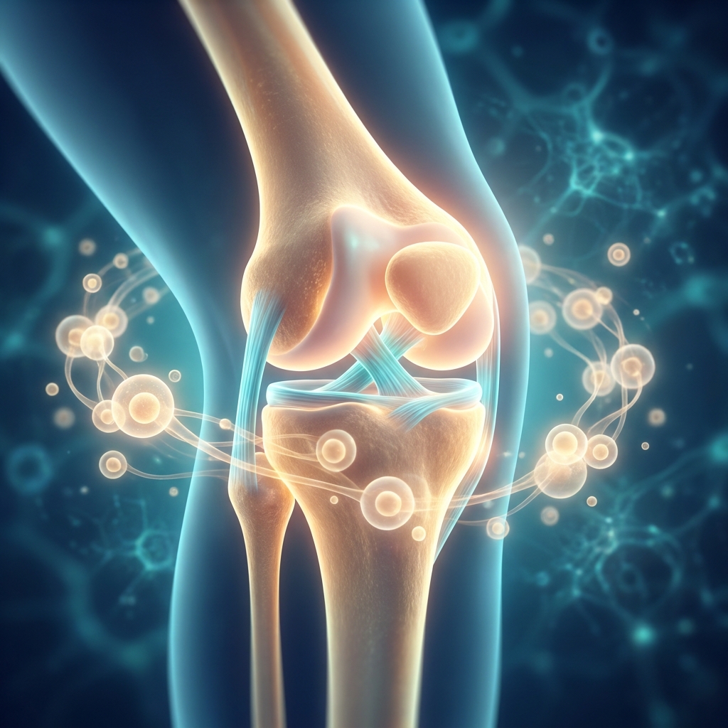
Regenerative Medicine for Ligament Injuries: The Biology-to-Treatment Framework That Explains Why Ligaments Fail to Heal and How Cellular Therapy Intervenes at Every Phase
Introduction: Why Ligament Injuries Remain One of Medicine’s Most Stubborn Healing Challenges
Ligament and tendon injuries are among the most common musculoskeletal problems worldwide. Together, they account for roughly 50% of the estimated 30-plus million musculoskeletal therapeutic procedures performed each year globally. For something so prevalent, ligament injuries share a frustrating trait: they heal poorly, slowly, and often incompletely.
The central paradox is this: the reason ligaments struggle to repair themselves has far less to do with how badly they are torn and far more to do with their fundamental biology. A ligament is built for strength, not for self-repair, and its tissue architecture actively works against the healing it requires.
This article takes a deliberately different approach from most discussions of regenerative medicine. Before explaining what therapies like platelet-rich plasma (PRP), mesenchymal stem cells (MSCs), bone marrow aspiration concentrate (BMAC), and exosomes actually do, it first explains why ligaments fail to heal on their own. Only with that biological foundation does the logic of each treatment become clear.
The discussion spans ligament injuries across multiple joints, including the knee, ankle, shoulder, wrist, and spine, reflecting the full scope of conditions treated at clinics like Unicorn Bioscience. Readers will follow the three-phase healing cascade and see exactly how each regenerative modality intervenes at specific stages of that process.
One note on regulation: as of 2026, the FDA has not approved stem cell, PRP, or exosome products specifically for orthopedic conditions. However, substantial clinical evidence supports their safety and efficacy when administered by qualified providers operating within FDA regulatory frameworks.
The Intrinsic Biology of Ligaments: Built for Strength, Not Self-Repair
Ligaments are dense, fibrous connective tissue structures that connect bone to bone. Their function is to stabilize joints and provide proprioceptive feedback, the sense of where a joint is in space. Their entire architecture is optimized for one purpose: withstanding mechanical load.
That optimization comes at a cost. Approximately 70 to 80% of the dry weight of a healthy ligament is type I collagen, the structural protein responsible for tensile stiffness and strength. This tightly packed, parallel-fiber composition gives a healthy ligament its mechanical integrity, but it leaves little room for the cellular and vascular machinery that drives repair.
Tissues that heal well, such as muscle and bone, are rich in cells and laced with blood vessels, giving them ready access to nutrients and reparative signals. Ligaments are the opposite, defined by two biological limitations:
- Hypovascularity: a limited blood supply that restricts delivery of oxygen, nutrients, and circulating reparative cells to an injury site.
- Hypocellularity: very few resident cells living within the tissue itself.
The sparse population of fibroblasts inside a ligament cannot mount a robust repair response on its own. The tissue depends heavily on outside signals and on cells migrating in from surrounding structures. As the next section explains, not all ligaments are created equal when it comes to healing potential.
Intra-Articular vs. Extra-Articular Ligaments: Why Location Determines Healing Fate
One of the most clinically important distinctions in ligament biology is anatomical location. Intra-articular ligaments, such as the anterior cruciate ligament (ACL) and posterior cruciate ligament (PCL), live inside the joint capsule and are bathed in synovial fluid. Extra-articular ligaments, such as the medial collateral ligament (MCL) of the knee and the lateral ligaments of the ankle, lie outside the capsule and have access to surrounding periligamentous vascular tissue.
This difference is decisive. Synovial fluid is hostile to healing because it prevents the formation of a fibrin clot. Normally, a fibrin clot bridges a wound gap and provides a structural template for cells to migrate across and deposit new collagen. Without that scaffold, the torn ends of an ACL cannot bridge the gap, and the body’s repair process stalls before it can begin.
The MCL tells the opposite story. Because it sits outside the joint capsule, a torn MCL allows a periligamentous hematoma to form. That hematoma becomes a fibrin scaffold that supports fibroblast infiltration and spontaneous healing. This is precisely why isolated MCL tears frequently heal with conservative care while ACL tears usually do not.
The same framework applies across the body. The glenohumeral ligaments of the shoulder, the scapholunate ligament of the wrist, the lateral ankle ligaments (ATFL and CFL), and the various spinal ligaments each occupy distinct vascular and anatomical environments that shape their healing capacity. The clinical takeaway is straightforward: intra-articular ligaments are far more dependent on regenerative interventions to compensate for the natural healing scaffold they lack.
Complicating matters further, between 54% and 71% of patients undergoing ACL surgery have at least one concomitant soft tissue injury, such as a meniscus, MCL, or PCL injury. This underscores why ligament care benefits from whole-joint thinking rather than treating a single structure in isolation.
The Type I vs. Type III Collagen Problem: Why Healed Ligaments Are Not the Same as Healthy Ligaments
Even when a ligament does heal, the repaired tissue is rarely equal to the original. The reason lies in collagen biology.
Healthy ligaments are dominated by type I collagen, with tightly packed, parallel fiber bundles that deliver high tensile strength. After an injury, however, the body rushes to close the wound by depositing type III collagen, which has a looser, more disorganized structure and inferior mechanical properties. This rapid deposition is biologically efficient, but the resulting tissue is mechanically weak.
The remodeling phase is supposed to gradually convert type III collagen back into well-organized type I collagen. In the absence of optimized biological signals, however, this conversion is incomplete. The result is healed tissue that behaves like scar rather than original ligament: weaker, less organized, and prone to re-injury.
This biological reality connects directly to surgical outcomes. Primary ACL reconstruction fails in roughly 3.2% to 11.1% of cases, and approximately 10% of reconstructed ACLs fail within 10 years, in part because graft remodeling and biological integration are imperfect. A comparative analysis found that biological autografts can fail at rates as low as 1.2%, while some synthetic grafts carry failure rates up to 33%, reinforcing that biology, not just hardware, drives durable healing.
Regenerative therapies, particularly PRP and MSCs, aim to modulate this collagen balance: favoring type III deposition early for rapid wound closure, then enhancing type I remodeling later to restore strength. This is one of the core biological targets of regenerative medicine.
The Epiligament: The Hidden Healing Reservoir That Changes Everything
A structure long overlooked in ligament biology is now emerging as one of the most important players in healing: the epiligament. This thin, metabolically active connective tissue layer envelops the outer surface of a ligament and serves as a critical healing reservoir.
Unlike the relatively barren interior of the ligament, the epiligament is rich in reparative fibroblasts, progenitor cells, blood vessels, and growth factors. After an injury, it serves as the primary source of cells that migrate into the damaged tissue.
Cutting-edge 2026 research suggests that differences in epiligament composition and vascularity may help explain why the ACL and MCL heal so differently. The ACL’s epiligament appears less vascular and less cellular than the MCL’s, compounding the disadvantage already imposed by the synovial environment.
The clinical implication is significant. Regenerative therapies that can activate, augment, or supplement the epiligament’s reparative cell population may unlock healing potential that the ligament’s interior cannot generate on its own. This biology-informed thinking distinguishes a sophisticated regenerative approach from conventional treatment and shapes how forward-looking providers like Unicorn Bioscience frame ligament care.
The Three-Phase Ligament Healing Cascade: A Biological Roadmap
Every regenerative therapy must navigate the same biological roadmap: a three-phase healing cascade. Understanding these phases is essential to understanding why specific treatments are applied at specific times. The phases overlap rather than proceed in strict sequence, and disruption of any one phase compromises the final outcome.
Phase 1: The Acute Inflammatory Phase (Days 0–7)
The moment a ligament is injured, vascular disruption triggers a hematoma, platelets activate, and a wave of pro-inflammatory cytokines (such as IL-1β and TNF-α) and chemokines recruits neutrophils and macrophages to the site.
Inflammation plays a dual role here. It is essential for clearing debris and initiating repair, but excessive or prolonged inflammation unleashes destructive matrix metalloproteinase (MMP) activity that degrades collagen and impairs healing. Platelets also release a cascade of growth factors, including PDGF, TGF-β, VEGF, and IGF-1, that begin recruiting and activating fibroblasts.
This makes Phase 1 a critical intervention window. Shifting inflammation toward a pro-healing M2 macrophage phenotype, rather than a destructive M1 phenotype, is one of the key mechanisms by which regenerative therapies improve outcomes. For intra-articular ligaments, the challenge is greater: synovial fluid dilutes growth factor concentrations and disrupts fibrin clot formation, making this phase far less productive for the ACL than for extra-articular ligaments.
Phase 2: The Proliferative and Regenerative Phase (Days 7–60)
In this phase, fibroblasts recruited from the epiligament, periligamentous tissue, and circulating progenitor cells proliferate and begin synthesizing new extracellular matrix, initially dominated by type III collagen.
Angiogenesis, the formation of new blood vessels driven by VEGF and other signals, is critical here for delivering oxygen, nutrients, and additional reparative cells. Unfortunately, this process is inherently limited in hypovascular ligament tissue. Growth factors orchestrate the work: TGF-β drives collagen synthesis, PDGF stimulates fibroblast proliferation, and IGF-1 supports cell survival and matrix production.
This phase is the primary target for MSC-based therapies, which can differentiate into ligament fibroblasts, secrete angiogenic growth factors, release anti-inflammatory cytokines, and promote matrix production simultaneously. The quality of collagen laid down here, including the type I to type III ratio and fiber alignment, largely determines the mechanical quality of the final tissue.
Phase 3: The Remodeling Phase (Months 2–24+)
The longest and most underappreciated phase is remodeling. Here, type III collagen is gradually replaced by type I, fiber bundles organize and align along lines of mechanical stress, and cellularity returns toward baseline. Full ligament maturation can take 12 to 24 months or longer, which is precisely why athletes who return to sport too early after ACL surgery face elevated re-injury risk.
Mechanical loading is essential to this phase. Controlled, progressive loading stimulates collagen alignment and cross-linking through mechanotransduction pathways, which is why structured rehabilitation is biologically necessary, not merely functional. Without adequate growth factor signaling, the type I conversion remains incomplete, leaving inferior scar tissue.
This phase is a target for sustained-release strategies: scaffolds, exosomes, and sequential PRP injections can extend bioactive signaling deep into remodeling to improve final tissue quality. The stakes are high, given that ACL reconstruction carries risks of donor-site morbidity, graft rejection, poor integration, and a high rate of post-traumatic osteoarthritis.
Regenerative Medicine for Ligament Injuries: How Each Therapy Intervenes in the Healing Cascade
With the biology established, each regenerative modality can be understood as a targeted intervention rather than a generic injection. The right choice, or combination, depends on the healing phase, the anatomical location (intra- versus extra-articular), the severity of the tear, and individual patient factors. Unicorn Bioscience’s personalized treatment planning accounts for inflammation levels, patient age, injury type and location, current medications, and personal health goals, reflecting exactly this biology-informed logic.
Platelet-Rich Plasma (PRP): Amplifying the Body’s Own Growth Factor Cascade
Its effect on collagen is particularly notable. By balancing TGF-β and PDGF activity, PRP favors type III collagen deposition early for rapid wound closure, then supports type I remodeling later to rebuild mechanical strength. In Phase 1, platelet-derived signals promote M2 macrophage polarization, dampening destructive inflammation while preserving pro-healing signals. In Phase 2, VEGF drives angiogenesis to counter hypovascularity, PDGF spurs fibroblast proliferation, and IGF-1 promotes cell survival.
Formulation matters. Leukocyte-rich PRP (LR-PRP) contains white blood cells that may amplify early inflammatory signals, potentially helpful in chronic injuries but possibly counterproductive in acute ones, while leukocyte-poor PRP (LP-PRP) carries a more anti-inflammatory profile. This is why preparation protocols influence outcomes. In vitro and animal studies consistently show improved biomechanical strength and histological quality with PRP, while human clinical evidence is encouraging but limited by protocol variability. PRP applies across joint regions, from intra-articular ACL and PCL (with imaging guidance to overcome synovial dilution) to the MCL, ankle ligaments, glenohumeral and scapholunate ligaments, and spinal ligaments.
Mesenchymal Stem Cells (MSCs) and BMAC: Cellular Regeneration Across All Healing Phases
MSCs are multipotent stromal cells derived from bone marrow (via BMAC), adipose tissue, or other sources, capable of differentiating into multiple connective tissue cell types, including ligament fibroblasts. BMAC is a minimally processed concentrate of the patient’s own bone marrow containing MSCs, hematopoietic progenitor cells, platelets, and growth factors.
MSCs act through several mechanisms simultaneously: (1) differentiation into fibroblasts to replenish hypocellular tissue; (2) secretion of anti-inflammatory cytokines such as IL-10 and TGF-β to calm Phase 1 inflammation; (3) release of angiogenic factors like VEGF to drive Phase 2 neovascularization; and (4) promotion of collagen synthesis in Phases 2 and 3. A 2015 prospective cohort study by Centeno and colleagues found that bone marrow-derived MSC injections for partial ACL tears improved knee stability and function, with MRI evidence of partial tissue regeneration.
MSCs are especially valuable for intra-articular ligaments because they can be delivered directly to the injury site under imaging guidance, compensating for the natural cell recruitment that synovial fluid prevents. Emerging evidence also suggests their primary mechanism may be paracrine signaling rather than direct differentiation, making their influence broader than once thought. At Unicorn Bioscience, all injections are administered using precision ultrasound and X-ray guidance, which is particularly important for accurately reaching intra-articular targets.
Exosomes and MSC-Derived Extracellular Vesicles: The Acellular Frontier of Ligament Regeneration
Their advantages over live cells are compelling: low immunogenicity, stable storage, consistent dosing, and the ability to cross biological barriers, including the synovial membrane. In ligament healing, exosomes modulate immunity (bone marrow MSC-derived exosomes promote M1-to-M2 macrophage polarization via miR-23a-3p), stimulate neovascularization through VEGF and angiogenic microRNAs, activate fibroblasts in the proliferative phase, and influence the balance between functional repair and fibrotic scar.
Their nano-scale size and low immunogenicity make exosomes especially well-suited for the synovial environment of intra-articular ligaments. This remains an emerging 2025 to 2026 frontier with growing preclinical evidence and early clinical exploration. Unicorn Bioscience offers exosome therapy as part of its cutting-edge portfolio, administered within FDA regulatory frameworks.
Biologic Scaffolds and the BEAR Technique: Structural Support Meets Biological Signaling
Even with optimal cells and growth factors, intra-articular ligaments lack the fibrin bridge that extra-articular ligaments form naturally. Without a structural scaffold, torn ends cannot reconnect. The Bridge-Enhanced ACL Repair (BEAR) technique, which is FDA-authorized, addresses this directly by placing a bioactive collagen scaffold saturated with the patient’s own blood between the torn ACL ends, supplying both the structural bridge and the biological signals needed for healing.
Across three BEAR clinical trials, the aggregate re-tear rate was 15%, with post-market registry data showing non-inferior patient-reported outcomes compared to ACL reconstruction and zero re-tears in some cohorts. The technique embodies the biology-first framework by artificially providing the scaffold the synovial environment otherwise prevents. Broader scaffold research, using collagen, hyaluronic acid, or silk-based materials, explores delivering PRP, stem cells, or growth factors across multiple joints. Industry momentum is also building: an active trial (NCT07129694) is evaluating CT-ACL001, a regenerative biological tissue ligament, in a randomized multicenter study with estimated completion in November 2027. Scaffolds complement rather than replace cellular therapies.
Combination Therapy: Why Multi-Modal Approaches May Outperform Single Modalities
Because ligament healing fails for multiple reasons simultaneously (hypovascularity, hypocellularity, collagen imbalance, absent scaffold, and a hostile synovial environment), no single modality addresses every barrier. This is the rationale for combination therapy.
PRP and MSCs complement each other: PRP supplies the immediate growth factor surge to launch Phases 1 and 2, while MSCs provide sustained cellular and paracrine support through Phases 2 and 3, including differentiation capacity PRP cannot offer. Exosomes can extend paracrine signaling beyond the lifespan of injected cells, and scaffolds combined with biologics tackle structural and biological deficits simultaneously. Unicorn Bioscience’s multi-modal menu (PRP, stem cell therapy, BMAC, and exosome therapy) allows customized combination protocols, though the evidence base for specific combinations is still developing, making experienced provider judgment essential.
Ligament Injuries Across the Body: Applying the Regenerative Framework Beyond the ACL
The biology-first framework is not limited to the knee. Hypovascularity, hypocellularity, collagen imbalance, and phase-specific healing apply to ligaments throughout the body, with anatomical variations. This mirrors the scope of care at Unicorn Bioscience across the shoulder, elbow, hand and wrist, spine, hip, knee, and ankle and foot.
Knee Ligaments: ACL, PCL, MCL, and LCL
The ACL remains the primary target for regenerative intervention given its intra-articular position, synovial environment, and absent fibrin scaffold. Its incidence is high in American football, basketball, soccer, and volleyball, with female athletes facing markedly higher rates; female basketball players show incidence up to 4.6 times that of male athletes. The MCL, being extra-articular, heals better, but severe Grade III tears or chronic instability may still benefit from PRP or MSC augmentation. The PCL, like the ACL, faces synovial challenges that make regenerative approaches relevant for partial tears. With 54 to 71% of ACL patients carrying concomitant injuries, whole-joint assessment is essential.
Ankle Ligaments: ATFL, CFL, and the Lateral Ligament Complex
Lateral ankle ligament injuries are among the most common sports injuries globally and are frequently undertreated, leading to chronic instability. These ligaments are extra-articular with better intrinsic healing, but recurrent sprains produce poor-quality scar tissue that benefits from regenerative augmentation. PRP can accelerate acute sprain healing and improve collagen quality in chronic cases, while MSC or BMAC therapy may suit severe injuries where conservative care has failed. Precision ultrasound guidance is particularly valuable given the complex lateral anatomy.
Shoulder Ligaments: Glenohumeral and Acromioclavicular
The glenohumeral ligaments are the primary static stabilizers of the shoulder, and injuries contribute to instability and recurrent dislocation. Because they are intra-articular, they face ACL-like synovial challenges, making regenerative options relevant. Acromioclavicular joint injuries (shoulder separations) are extra-articular with better healing potential, though high-grade separations may benefit from PRP. These approaches are attractive for active individuals seeking to avoid lengthy surgical recovery, and imaging guidance is essential given nearby neurovascular structures.
Wrist and Hand Ligaments: Scapholunate and Beyond
The scapholunate ligament is one of the most clinically significant wrist ligaments; untreated injury leads to carpal instability and progressive arthritis. Wrist ligaments are small, complex, and poorly vascularized, making them weak intrinsic healers and strong candidates for augmentation. PRP can support partial scapholunate tears, particularly for patients exploring non-surgical options. Precision imaging guidance is critical given the small target size.
Spinal Ligaments: Interspinous, Supraspinous, and Ligamentum Flavum
Spinal ligaments contribute to vertebral stability, and their injury or degeneration can drive chronic back pain that conventional imaging often underestimates. PRP and MSC-based therapies can be applied in the context of chronic low back pain or post-surgical instability, an area of growing clinical interest. Given proximity to neural structures, precise imaging guidance and experienced providers are essential.
Who Is a Candidate for Regenerative Ligament Therapy? Understanding the Patient Journey
Several patient profiles tend to benefit from regenerative ligament therapy:
- Partial-tear patients (Grade I–II) who have not responded adequately to rest, physical therapy, and bracing and want to optimize healing before considering surgery.
- Post-surgical augmentation candidates seeking to enhance graft integration, reduce inflammation, and improve remodeling.
- Failed-surgery candidates who experienced re-tear or poor outcomes after reconstruction and want a second-chance option for partial healing or improved stability.
- Athletes in high-demand sports, including the disproportionately affected population of female athletes, for whom faster, biology-driven recovery is a priority.
Personalized planning at Unicorn Bioscience accounts for inflammation levels, age, injury type and location, current medications, and personal goals, ensuring biology-informed rather than one-size-fits-all selection. Not every patient is a candidate for every modality, so thorough clinical assessment including imaging is essential. Virtual and in-person consultations are available across the company’s eight locations in Texas, Florida, and New York.
The Current Evidence Landscape and What Patients Should Know
An honest assessment of the evidence matters. In vitro and animal studies consistently show improved biomechanical and histological outcomes with PRP and MSC therapies, while human clinical evidence is growing but still limited by small samples, heterogeneous preparation protocols, and a shortage of large randomized controlled trials. Key challenges include standardizing cell procurement and PRP preparation, controlling cytokine and gene delivery, revascularizing avascular tissue, and conducting well-powered trials.
The trajectory is positive. There are 224 clinical trials globally investigating stem cell therapies for osteoarthritis, a major Phase III trial funded with $140 million was announced in January 2026, and the CT-ACL001 randomized multicenter trial is actively progressing. As of 2026, the FDA has not approved stem cell, PRP, or exosome products specifically for orthopedic conditions, but substantial evidence supports their safety and efficacy when administered by qualified providers within FDA regulatory frameworks. Set against surgical alternatives, with their failure rates of 3.2 to 11.1%, donor-site morbidity, graft rejection, and post-traumatic osteoarthritis risk, regenerative approaches deserve serious consideration. The goal is not to eliminate surgery but to provide biology-informed, minimally invasive alternatives or adjuncts for appropriate candidates.
Conclusion: From Biology to Treatment, a New Standard for Ligament Care
Ligaments do not fail to heal because the body lacks the will to repair. They fail because their intrinsic biology, defined by hypovascularity, hypocellularity, type III collagen dominance, and (for intra-articular ligaments) a hostile synovial environment, creates specific and addressable biological deficits.
The emerging science of the epiligament as a healing reservoir reframes our understanding of repair potential and explains why targeted regenerative delivery to that biological interface matters. The phase-specific framework brings the picture together: PRP amplifies growth factor signaling across all phases, MSCs and BMAC provide cellular and paracrine support during proliferation and remodeling, exosomes deliver acellular immune modulation and angiogenic stimulation, and scaffolds resolve the structural deficit unique to intra-articular ligaments.
This framework applies throughout the body, from knee and ankle to shoulder, wrist, and spine. As the clinical evidence base matures and protocols standardize, regenerative medicine for ligament injuries is positioned to become a cornerstone of non-surgical orthopedic care. Unicorn Bioscience embodies this direction, combining cutting-edge cellular therapies with precision imaging guidance and personalized, biology-informed planning across eight locations in Texas, Florida, and New York.
Take the Next Step: Explore Regenerative Options for Your Ligament Injury
For anyone struggling with a ligament injury, whether a recent tear, ongoing instability, or a disappointing surgical outcome, the most empowering first step is understanding the biology behind the problem. The second step is finding a provider who treats that biology accordingly.
Unicorn Bioscience offers both virtual and in-person consultations, making expert regenerative care accessible to patients across Texas (Austin, Dallas, El Paso, Fort Worth, Houston, and San Antonio), Florida (Boca Raton), and New York (Manhattan). Qualified candidates may even receive same-day treatment, shortening the gap between consultation and intervention.
To schedule a consultation and receive a personalized treatment assessment, contact Unicorn Bioscience at (737) 347-0446 or visit unicornbioscience.com. Every patient’s biology is unique, and their treatment should be too.
Schedule Your Consultation Today!
