
Achy Pain Behind Knee: The Sitting-to-Serious Risk Ladder That Tells You Exactly When to Act
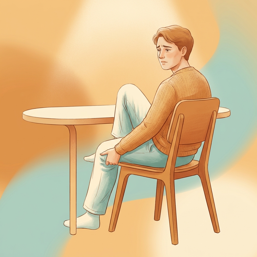
Achy Pain Behind Knee: The Sitting-to-Serious Risk Ladder That Tells You Exactly When to Act
Introduction: That Dull, Achy Pain Behind Your Knee Is Telling You Something
That persistent, nagging ache behind the knee is different from a sudden, sharp pain after a twist or fall. The achy pain behind the knee that many people experience carries its own diagnostic significance, signaling chronic tissue stress, inflammation, or fluid accumulation rather than an acute structural injury. This distinction matters because it helps narrow down the likely causes and determines the appropriate response.
Knee pain is remarkably common. According to data from the American Academy of Family Physicians, approximately 25% of adults experience frequent knee pain, and its prevalence has increased nearly 65% over the past 20 years. The back of the knee, known clinically as the popliteal fossa, is a complex anatomical region housing tendons, muscles, nerves, blood vessels, and bursae. This complexity explains why so many different conditions can produce aching in this specific location.
This article presents a “risk ladder” framework that organizes the causes of posterior knee aching from the most benign (sedentary lifestyle-driven tightness) to the most serious (deep vein thrombosis). Readers can locate themselves on this spectrum and understand exactly when to act. While most causes are manageable with conservative care, a few require urgent medical attention. Knowing the difference is the goal of this guide.
What “Achy” Actually Means and Why the Pain Quality Matters
The clinical distinction between achy, dull pain and sharp, acute pain is significant. Achy pain typically signals chronic or subacute tissue stress, inflammation, or fluid accumulation rather than acute structural failure such as a torn ligament.
Key characteristics of achy posterior knee pain include:
- Persistent discomfort that lingers throughout the day
- Worsening after prolonged sitting or activity
- A sensation of pressure, tightness, or fullness
- Gradual onset over days or weeks
- Often bilateral or affecting both knees
In contrast, red-flag pain qualities signal a different, more urgent category. These include sudden sharp pain, pain that wakes a person from sleep, pain accompanied by immediate swelling, or an inability to bear weight.
The “achy” quality narrows the differential diagnosis toward conditions such as Baker’s cyst, hamstring tendinopathy, osteoarthritis, bursitis, and early meniscal involvement. The precise location within the back of the knee (medial, lateral, or central) can further refine the likely cause.
A Quick Guide: What Does Your Achy Knee Pain Feel Like?
Before diving into the full risk ladder, this symptom-mapping framework helps readers self-identify their likely cause:
- Aching after sitting for hours at a desk: Likely hamstring tightness or early tendinopathy
- Aching with a visible or palpable lump behind the knee: Likely Baker’s cyst
- Aching that worsens when bending or squatting, especially after age 40: Likely osteoarthritis or posterior meniscus involvement
- Aching on the inner back of the knee in older adults: Consider semimembranosus tendinopathy
- Aching accompanied by swelling, warmth, and redness that worsens over days: Urgent: possible DVT
- Aching behind the knee alongside lower back pain or leg tingling: Consider spinal nerve referral
This guide provides orientation only. A healthcare provider should confirm any diagnosis.
The Sitting-to-Serious Risk Ladder: 7 Causes of Achy Pain Behind the Knee
The risk ladder organizes causes from lowest urgency (benign, self-treatable) to highest urgency (requiring immediate medical attention). “Lower on the ladder” does not mean “less painful.” It means less medically dangerous. Each rung includes who is most at risk, what the aching feels like, and what to do about it.
Rung 1: Hamstring Tightness and Sedentary Lifestyle (Lowest Risk)
Tight or weak hamstrings, often caused by prolonged sitting, pull on their insertion points at the back of the knee, generating a dull, diffuse ache. Research indicates that sitting for over four hours is associated with a high risk of significant hamstring tightness and posterior knee pain.
The typical sufferer is a desk worker, driver, frequent flyer, or anyone with a predominantly sedentary lifestyle. This is muscular tightness, not structural tissue damage, making it the most benign and reversible cause on the ladder.
This “sitting-too-long” trigger represents a modern, underappreciated cause that many healthcare resources overlook. First-line responses include standing breaks every 30 to 60 minutes, hamstring stretching, and progressive strengthening exercises.
Rung 2: Hamstring Tendinopathy (Low-Moderate Risk)
Distal hamstring tendinopathy involves degeneration or micro-tearing of the hamstring tendon where it attaches near the back of the knee. According to Physiopedia, the main symptoms include an “achy feeling” and stiffness that typically set in gradually during or after activity.
Unlike simple tightness, the pain is more localized, often tender to touch at the tendon insertion, and worsens during or after activity rather than resolving with movement. Those most at risk include runners, cyclists, athletes, and active individuals. Sedentary people who suddenly increase activity are also vulnerable.
Semimembranosus tendinopathy (SMT) is a specific, underdiagnosed subtype causing chronic posteromedial knee aching. It is more common in older patients and often missed in clinical practice.
Treatment approaches include activity modification, eccentric strengthening exercises, and physical therapy. Persistent cases may benefit from regenerative options such as PRP therapy for hamstring tendinopathy, which is commonly used for tendinopathy.
Rung 3: Baker’s Cyst (Popliteal Cyst) (Low-Moderate Risk)
A Baker’s cyst is a fluid-filled sac that forms in the popliteal fossa when excess joint fluid is pushed into a natural pocket behind the knee. According to StatPearls, Baker’s cysts occur most commonly in adults aged 35 to 70 and are strongly associated with underlying osteoarthritis or meniscal tears.
The typical presentation includes a soft, sometimes visible lump behind the knee accompanied by a dull ache or pressure sensation, often worse after prolonged standing or activity. These cysts have been found in 4.7% to 37% of adults with asymptomatic knees on imaging.
Crucially, Baker’s cysts are almost always a symptom of an underlying joint problem, not a standalone condition. Treating the root cause is essential.
When a cyst ruptures, fluid leaks into the calf, causing sudden swelling, pain, and warmth. This is called pseudo-thrombophlebitis and can look exactly like DVT, setting up a critical diagnostic challenge addressed later in this article.
Rung 4: Osteoarthritis and Posterior Meniscus Tears (Moderate Risk)
Osteoarthritis causes posterior knee aching through cartilage breakdown, joint inflammation, and bone spur formation. This condition affects over 365 million people globally and is the leading cause of chronic knee pain worldwide. Prevalence spikes sharply after age 45, with up to 50% of individuals over 75 reporting knee discomfort.
Tears in the posterior horn of the meniscus cause aching behind the knee, particularly with twisting, squatting, or deep bending. Patients often describe a “catching” ache. Osteoarthritis and posterior meniscal tears frequently coexist, especially in adults over 50.
A landmark 2025 study published in the New England Journal of Medicine (the TeMPO trial) confirmed that all participants with knee osteoarthritis and meniscal tear improved substantially with exercise. Those who also saw a physical therapist reported slightly greater pain relief at 6 and 12 months. Additionally, a 2025 BMJ network meta-analysis of 217 randomized trials found aerobic exercise consistently ranked as the most effective intervention for knee OA pain.
Treatment options include exercise therapy, physical therapy, NSAIDs, and weight management. Regenerative medicine options such as PRP, stem cell therapy, and hyaluronic acid injections are used as non-surgical alternatives to knee replacement for appropriate candidates.
Rung 5: Spinal Nerve Referral (S1/S2 Radiculopathy) (Moderate Risk, Often Overlooked)
Compression of the S1 or S2 spinal nerve roots in the lumbar spine can refer pain down the back of the leg and concentrate it behind the knee, even when the knee itself is structurally normal.
Distinguishing features include posterior knee aching accompanied by lower back pain, buttock pain, or tingling and numbness down the leg. The pain may not change with knee-specific movements.
This represents a commonly missed diagnosis. Patients and even some clinicians focus on the knee and miss the spinal origin. Adults with a history of back problems, disc herniation, or degenerative spine disease are most at risk. Treating the knee will not resolve this pain; the spine must be addressed.
Rung 6: Popliteal Bursitis and PCL Injury (Moderate-High Risk)
Popliteal bursitis involves inflammation of one of the bursae in the popliteal fossa, producing localized aching and tenderness, often from overuse or direct trauma.
The posterior cruciate ligament (PCL) stabilizes the knee from behind. Injury, often from a direct blow to the front of the knee, causes posterior aching, instability, and swelling. There is usually a clear mechanism of injury such as a fall, car accident, or sports contact. The aching is accompanied by a sense of instability or giving way.
PCL injuries are less common than ACL injuries but are sometimes underdiagnosed because the knee may still feel functional. Treatment often responds well to conservative rehabilitation, though severe cases may require surgical evaluation.
Rung 7: Deep Vein Thrombosis (DVT) (Highest Risk, Requires Urgent Action)
DVT occurs when a blood clot forms in the popliteal vein behind the knee or in deeper leg veins. This is a medical emergency because the clot can travel to the lungs, causing a pulmonary embolism.
According to the Mayo Clinic, DVT presents as aching or throbbing pain behind the knee that worsens over time and does not improve with rest. It is accompanied by swelling, warmth, and redness in the calf or behind the knee.
DVT may go unnoticed in approximately 30 to 40% of cases, making awareness of warning signs critical. Risk factors include prolonged sitting (long flights, car rides, desk work), recent surgery, immobility, pregnancy, clotting disorders, and older age.
If DVT is suspected, seek emergency medical care immediately. Do not massage the area.
Baker’s Cyst vs. DVT: The Comparison That Could Save Your Life
This is the most anxiety-provoking diagnostic confusion patients face. Both conditions can cause swelling, aching, and discomfort behind the knee and in the calf, but they require completely opposite responses. A ruptured Baker’s cyst can closely mimic deep vein thrombosis, often leading to diagnostic uncertainty and delayed management.
| Feature | Baker’s Cyst | DVT |
|---|---|---|
| Onset | Gradual | Progressive worsening |
| Feel | Soft lump | Diffuse swelling |
| Skin changes | Usually none | Warmth and redness |
| Response to rest | Often improves | Does not improve |
| Associated symptoms | None | Possible shortness of breath |
| Action | Conservative care | Emergency care |
When a Baker’s cyst ruptures, fluid tracks into the calf and mimics DVT almost perfectly. Even experienced clinicians can be fooled, and ultrasound is often required to differentiate.
The clear action rule: If there is any doubt, especially if pain is worsening, the calf is swollen and warm, or DVT risk factors are present, go to the emergency room or urgent care. Do not self-diagnose and wait.
The Sitting-Too-Long Problem: Why a Desk Job May Be Behind the Knee Ache
Prolonged sitting shortens and tightens the hamstrings, compresses the popliteal fossa, reduces circulation, and increases fluid accumulation behind the knee. Sitting for over four hours is associated with a high risk of significant hamstring tightness and posterior knee pain. Prolonged sitting is also a recognized DVT risk factor.
The typical pattern involves an ache that develops gradually over the workday, is worst after rising from a chair, and may ease with movement, only to return after the next long sitting period.
Practical interventions for desk workers include:
- Standing breaks every 30 to 60 minutes
- Seated hamstring stretches
- Proper desk ergonomics (chair height with hips and knees at 90 degrees, feet flat on floor)
- Avoiding crossing legs
- Brief walking intervals
Chronic hamstring tightness from prolonged sitting can progress to tendinopathy if unaddressed. If the ache is accompanied by any swelling or warmth after a very long period of immobility, DVT must be considered.
When to See a Doctor: Your Action Checklist
Urgent/Emergency (go now):
- Sudden severe swelling behind the knee or in the calf
- Warmth, redness, and pain worsening over hours or days
- Shortness of breath or chest pain alongside knee symptoms
- Fever with joint swelling
- Inability to bear weight after an injury
See a doctor within a few days:
- Aching persisting for more than one to two weeks without improvement
- A visible or growing lump behind the knee
- Aching accompanied by lower back pain or leg tingling
- Pain that woke the person from sleep
- Any posterior knee pain after recent surgery, long travel, or prolonged immobility
Monitor at home (but reassess if no improvement):
- Mild aching that began after increased activity or prolonged sitting
- Aching that responds to rest, ice, and stretching
- No swelling, redness, or warmth
Regarding imaging: MRI is appropriate for suspected meniscal or ligament involvement; ultrasound-guided evaluation helps differentiate Baker’s cyst from DVT; X-ray is useful for suspected arthritis or bony changes.
Treatment Options: From Home Remedies to Regenerative Medicine
At-Home First-Line Care
The RICE method (Rest, Ice, Compression, Elevation) is the standard first response for most benign causes of achy posterior knee pain. NSAIDs such as ibuprofen or naproxen can help with inflammation and pain management.
Gentle stretching and strengthening, including hamstring stretches, calf stretches, and progressive strengthening exercises, are evidence-based first-line interventions. The 2025 NEJM TeMPO trial and 2025 BMJ aerobic exercise meta-analysis confirm that exercise, particularly aerobic exercise, is consistently the most effective intervention for knee OA pain and meniscal conditions.
Physical Therapy and Medical Management
Physical therapy addresses the root biomechanical cause, not just the symptom. This is particularly important for tendinopathy, PCL injury, and post-meniscal conditions.
Corticosteroid injections may be appropriate for Baker’s cysts and bursitis in some cases, though with limitations on frequency and long-term use. Hyaluronic acid injections provide joint lubrication and pain relief for osteoarthritis.
For Baker’s cysts, treating the underlying arthritis or meniscal tear is more effective than treating the cyst itself.
Regenerative Medicine: A Non-Surgical Option for Appropriate Causes
For patients who have not responded to conservative care and want to avoid surgery, regenerative medicine offers promising alternatives.
PRP (Platelet-Rich Plasma) therapy uses the patient’s own blood to deliver concentrated growth factors to damaged tissue. It is particularly well-suited for tendinopathy, osteoarthritis, and meniscal conditions. Stem cell therapy and BMAC (Bone Marrow Aspiration Concentrate) promote tissue repair and may reduce the need for joint replacement. Research suggests that up to 80% of patients told they need knee replacement may not actually require surgery.
All regenerative injections should be administered with ultrasound or imaging guidance to ensure accurate placement. As of 2026, the FDA has not approved stem cell, PRP, or exosome products specifically for orthopedic conditions, but substantial clinical evidence supports safety and efficacy when administered by qualified providers within FDA regulatory frameworks. Currently, 224 clinical trials globally are investigating stem cell therapies for osteoarthritis, and a major $140 million Phase III trial was announced in January 2026.
Regenerative medicine is appropriate for persistent tendinopathy, osteoarthritis, meniscal conditions, and patients seeking surgery alternatives. It is not appropriate for DVT or spinal referral pain.
Prevention: Keeping the Ache From Coming Back
Hamstring strengthening protocol: Progressive eccentric exercises such as Nordic curls and Romanian deadlifts are the most evidence-based approach to preventing hamstring tendinopathy.
Desk ergonomics for sedentary workers: Chair height should position hips and knees at 90 degrees, feet flat on floor. Avoiding crossed legs and incorporating standing desk intervals are also recommended.
Activity modification for runners and cyclists: Gradual mileage increases, proper bike fit, and avoiding sudden training load spikes reduce injury risk.
Weight management: Every extra pound adds approximately five extra pounds of pressure on the kneecap when climbing stairs. Weight reduction meaningfully reduces posterior knee stress.
DVT prevention during travel: Regular movement, hydration, and compression socks for long flights or drives help prevent clot formation.
Regular low-impact aerobic exercise: The 2025 BMJ meta-analysis shows aerobic exercise is the most effective long-term strategy for knee OA pain prevention and management.
Adults over 45 should be proactive about joint health, as prevalence of knee pain spikes significantly in this age group.
Conclusion: Reading the Knee’s Signals and Acting at the Right Time
The “achy” quality of posterior knee pain is a meaningful signal. It narrows the likely causes and points toward conditions that are usually manageable, though occasionally urgent.
The risk ladder framework spans from sedentary lifestyle-driven hamstring tightness at the benign end, through Baker’s cyst, tendinopathy, osteoarthritis, and spinal referral, to DVT at the serious end. Knowing where a person sits on this ladder determines exactly when to act.
The Baker’s cyst versus DVT distinction deserves special attention. When in doubt, seeking medical evaluation is the appropriate course of action. This is the one comparison where self-diagnosis can have serious consequences.
Most causes of achy pain behind the knee respond well to conservative care, exercise, and, for persistent cases, regenerative medicine options that can help patients avoid surgery. Understanding the pain is the first step to resolving it.
Ready to Address Achy Knee Pain? Explore Options With Unicorn Bioscience
For patients whose posterior knee pain has persisted beyond home care or who are exploring non-surgical options, Unicorn Bioscience offers regenerative medicine alternatives including PRP, stem cell therapy, BMAC, exosomes, and hyaluronic acid injections. All procedures use precision imaging-guided injections, with personalized treatment planning and same-day treatment availability for qualified candidates.
Unicorn Bioscience operates eight locations across Texas (Austin, Dallas, El Paso, Fort Worth, Houston, San Antonio), Florida (Boca Raton), and New York (Manhattan). Virtual consultation options are also available.
To discuss specific posterior knee pain concerns and explore whether regenerative medicine is appropriate, schedule a consultation by calling (737) 347-0446 or visiting unicornbioscience.com.
Schedule Your Consultation Today!
