
PRP Injection for Hip Arthritis: The Deep-Joint Delivery Framework That Explains Why the Hip Demands a Different Protocol Than Every Other Joint
PRP Injection for Hip Arthritis: The Deep-Joint Delivery Framework That Explains Why the Hip Demands a Different Protocol Than Every Other Joint
Introduction: Why Hip Arthritis Deserves Its Own PRP Conversation
Osteoarthritis is one of the most widespread sources of pain and disability on the planet. In 2021, approximately 607 million people worldwide were living with some form of the disease, and the trajectory is steepening. According to the Global Burden of Disease Study 2021, hip osteoarthritis cases alone are projected to increase by 78.6% by 2050. That is not a slow drift; it is a tidal wave bearing down on health systems already strained by demand for joint replacements.
Despite this, most patient-facing content about platelet-rich plasma (PRP) injections suffers from a stubborn problem: it either leans almost entirely on knee arthritis data or treats every joint in the body as interchangeable. That is a clinical oversimplification, and it can quietly mislead patients into the wrong expectations and, sometimes, the wrong procedure.
The thesis of this article is straightforward. The hip is not the knee. As a deep ball-and-socket joint buried beneath thick muscle and a fibrous capsule, the hip presents biological, procedural, and clinical realities that demand a fundamentally different PRP protocol. To make sense of those differences, this article introduces the Deep-Joint Delivery Framework: five pillars covering hip-specific biology, imaging guidance requirements, hip-specific clinical evidence, PRP formulation distinctions, and advanced combined techniques.
The goal here is not to replace a physician consultation. It is to help patients with hip arthritis ask sharper questions, recognize what separates a high-quality hip PRP procedure from a careless one, and understand whether they might be a candidate at all.
Understanding Hip Osteoarthritis: The Joint That Breaks the Rules
Hip osteoarthritis is a chronic degenerative joint disease, far more than simple “wear and tear.” The condition involves the progressive breakdown of cartilage, the formation of osteophytes (bone spurs), and changes deep within the subchondral bone that sits beneath the cartilage surface.
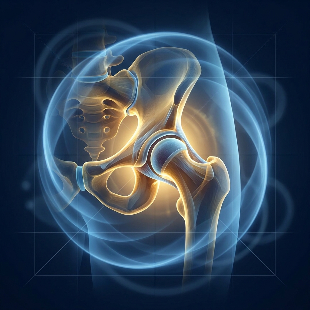
What makes the hip structurally unique is its architecture. The femoral head (the “ball”) seats deep within the acetabulum (the “socket”), and the whole assembly is wrapped in thick musculature, a dense fibrous joint capsule, and a cartilage rim called the labrum. These layers create access challenges that simply do not exist in more superficial joints.
Prevalence also varies dramatically by region. A 2023 systematic review of 31 studies covering over 326,000 participants found hip OA proportions ranging from 3.3% in East Asia to 9.5% in high-income North America.
Clinicians classify hip OA severity using the Kellgren-Lawrence (KL) grading scale, which runs from Grade 1 (minimal) to Grade 4 (severe, bone-on-bone). That framework will reappear throughout this article, because candidacy for PRP hinges heavily on where a patient falls on that scale. Hip OA is a leading cause of pain, lost function, and reduced quality of life worldwide, and demand for both surgical and non-surgical orthopedic solutions is accelerating.
The Deep-Joint Delivery Framework: Why the Hip Is Not the Knee
The Deep-Joint Delivery Framework organizes the hip PRP conversation around five dimensions: biology, access, evidence, formulation, and technique.
Consider joint depth alone. The knee joint space is relatively accessible, sitting close to the surface with palpable landmarks. The shoulder’s glenohumeral joint is moderately deep. The hip, by contrast, sits beneath layers of muscle and fat at a depth that routinely requires spinal-length needles to reach.
This is not an academic distinction. Conflating hip PRP protocols with knee PRP protocols has real consequences for whether the injected growth factors ever reach the intra-articular space where they are supposed to work. The following sections unpack each of the five pillars in turn.
Framework Pillar 1: The Hip-Specific Biology of PRP
At its foundation, PRP is autologous, meaning it comes from the patient’s own body. A small blood sample, typically between 15 and 150 mL, is drawn from the arm and centrifuged to concentrate platelets to roughly two to five times above baseline. The resulting plasma is then injected back into the joint, which minimizes any risk of allergy or rejection.
The therapeutic action comes from the growth factors that concentrated platelets release: TGF-β (transforming growth factor-beta), PDGF (platelet-derived growth factor), VEGF (vascular endothelial growth factor), and IGF-1 (insulin-like growth factor-1). These signaling molecules perform several functions simultaneously.
First, they modulate the NF-κB pathway. In an arthritic hip, NF-κB signaling drives chronic inflammation and the production of cartilage-degrading enzymes. PRP growth factors help suppress this pathway, reducing the catabolic, tissue-destroying environment inside the joint.
Second, PRP stimulates synovial hyaluronic acid production. It signals the synoviocytes lining the joint to manufacture more of the body’s own hyaluronic acid, improving lubrication from the inside out. A 2025 Frontiers in Bioengineering meta-analysis supports this lubrication-enhancing mechanism.
Collectively, these effects push the joint environment away from breakdown and toward repair and maintenance. That is a fundamentally different goal than corticosteroids, which primarily block pain rather than address the underlying biology. Understanding how PRP’s inflammation response works helps clarify why it may also stimulate chondrocyte proliferation, which can help slow cartilage degeneration. It is critical, however, to set realistic expectations: PRP is not a cartilage regeneration cure.
Framework Pillar 2: Why Ultrasound Guidance Is Non-Negotiable for the Hip
The hip is one of the deepest joints in the body. Its intra-articular space sits beneath the capsule, surrounded by the iliopsoas muscle, the femoral neurovascular bundle, and significant adipose tissue. Reaching it accurately is a genuine technical challenge.
The clinical implication is direct: blind, landmark-guided injections that might be acceptable in a superficial joint carry a meaningful risk of extra-articular placement in the hip. When that happens, the PRP never reaches the joint space, and the entire procedure is wasted.
The standard of care relies on real-time ultrasound, which allows the physician to guide a 20 to 22 gauge spinal needle (typically 90 to 120 mm in length) precisely into the joint space. Ultrasound enables dynamic visualization of the needle tip, confirmation of intra-articular placement before injection, and safe avoidance of the femoral nerve and artery.
A knee injection can often be performed accurately without imaging because of its superficiality. The hip offers no such margin. For more advanced techniques, fluoroscopic (X-ray) guidance comes into play, as discussed in Pillar 5. Unicorn Bioscience administers all of its injections using advanced imaging guidance, including ultrasound and X-ray, which is consistent with this standard of care.
Framework Pillar 3: What the Hip-Specific Clinical Evidence Actually Shows
The first honest point is that most published PRP outcome data comes from knee studies. Extrapolating those findings to the hip is scientifically shaky given the anatomical and biomechanical differences. Fortunately, the hip-specific literature is growing.
A 2024 systematic review of 5 RCTs published in Cureus found that every included study demonstrated significant pain reduction and functional improvement with PRP for hip OA, with no major adverse events. A separate meta-analysis of 7 RCTs covering 478 participants compared PRP against hyaluronic acid and found PRP notably more effective at reducing pain at the 6-month mark, outperforming HA through the 6 to 12 month mid-term window.
A real-world image-guided hip PRP study of 36 patients showed durable benefit: 67% reported greater than 50% improvement in pain at 3 months, and 58% maintained that improvement at 6 months. A 2022 RCT in BMC Musculoskeletal Disorders involving 105 patients with KL Grade 2 to 3 disease found PRP and PRP+HA effects lasted longer and outperformed HA alone on function, disability, and activities of daily living. A 2025 double-blind RCT on hip OA with developmental dysplasia found the PRP group achieved significantly higher pain-score improvement than the HA group over 24 weeks using three ultrasound-guided injections.
Honesty requires presenting the counterpoint. A 2024 double-blind RCT of 60 patients found PRP showed no significant advantage over saline placebo on activity pain scores at 6 months. The debate in the literature is real.
One detail helps reconcile the mixed results. Recent 2024 to 2025 meta-analyses show PRP reaches the Minimal Clinically Important Difference on the WOMAC scale at 1, 3, 6, and 12 months, but only when platelet concentration exceeds 1 million platelets per microliter. The evidence is generally positive, but preparation quality matters enormously, which leads directly to Pillar 4.
Framework Pillar 4: LR-PRP vs. LP-PRP, The Formulation Decision That Changes Everything for Hip OA
Not all PRP is the same. The leukocyte (white blood cell) content of the final preparation is one of the most clinically significant variables in the entire field, and one of the least discussed in patient-facing material.
Leukocyte-Rich PRP (LR-PRP) contains a high concentration of white blood cells alongside platelets. It has historically been used in tendon and soft tissue applications where a controlled inflammatory response may aid healing. Leukocyte-Poor PRP (LP-PRP) has most of those white blood cells removed, producing a more anti-inflammatory cytokine profile.
For hip OA, LP-PRP is generally preferred. The arthritic joint is already in a state of chronic inflammation, and introducing leukocyte-rich material can amplify that inflammatory response, causing increased post-injection pain and swelling. One meta-analysis found LR-PRP carried an odds ratio of 3.3 for adverse effects compared to LP-PRP, a clinically meaningful difference for patients already managing chronic hip pain.
A 2026 systematic review, accepted in February 2026, examined 11 studies on LR-PRP versus LP-PRP for OA and confirmed that leukocyte concentration significantly influences both efficacy and side-effect profiles. The practical takeaway for patients: asking a provider whether they use LP-PRP or LR-PRP for hip OA, and why, is a meaningful quality indicator. The broader lack of standardization across centrifuge speed, platelet concentration, leukocyte content, injection volume, and number of injections remains the field’s biggest obstacle.
Framework Pillar 5: The Combined Intra-Articular Plus Intraosseous Technique for Advanced Cases
For patients with more advanced hip OA who may not get sufficient relief from a standard injection, the literature describes a combined intra-articular plus intraosseous PRP infiltration technique.
The rationale is anatomical. In advanced hip OA, the pathology extends beyond the cartilage surface into the subchondral bone. A standard intra-articular injection delivers PRP to the synovial fluid and cartilage surface but never reaches that bone layer. The combined technique uses ultrasound guidance for the intra-articular component and adds fluoroscopic guidance to deliver PRP directly into the subchondral bone.
The biological goal is comprehensive coverage: growth factors reaching the synovial fluid, articular cartilage, and subchondral bone simultaneously, addressing the full spectrum of tissue involved. A PMC case series describes this approach for severe disease.
This is not a first-line treatment. It is reserved for more advanced cases where standard injection has proven insufficient or imaging shows significant subchondral involvement. It also demands additional equipment and procedural expertise, which is one more reason provider selection matters for the hip. Understanding the benefits of X-ray guided joint injection helps clarify why fluoroscopic guidance is essential for this advanced technique.
Who Is a Good Candidate for PRP Hip Injections?
The optimal candidate typically has mild-to-moderate hip OA (KL Grades 1 to 3), preserved joint space on imaging, falls roughly in the 49 to 68 age range seen in most studied cohorts, has a BMI under 35, and has already failed conservative treatments such as NSAIDs, physical therapy, and corticosteroid injections.
Expectations must be realistic. PRP is not a cure and does not regenerate lost cartilage. The goal is meaningful pain reduction, improved function, and potentially slowing progression, not reversal.
Patients with severe bone-on-bone disease (KL Grade 4) are generally poor candidates for PRP alone; total hip replacement is usually the more appropriate intervention at that stage. Other factors that may rule out PRP include active infection, bleeding disorders, anticoagulant use, active cancer, and certain autoimmune conditions. A BMI above 40 may reduce both procedural accuracy and outcomes. Determining candidacy requires a thorough clinical evaluation that includes an imaging review, which is why a physician consultation is essential.
How the Procedure Is Performed: What Patients Should Expect
The process is methodical. Blood is drawn from the arm (typically 15 to 150 mL), centrifuged to concentrate the platelets, and the resulting PRP is prepared for injection.
For the injection itself, the patient is positioned on the treatment table. Ultrasound identifies the hip joint space and the surrounding neurovascular structures. A spinal-length needle (20 to 22 gauge, 90 to 120 mm) is advanced under real-time visualization into the intra-articular space, and the PRP is injected only after correct placement is confirmed.
Most hip protocols involve a series of injections, commonly two to three spaced weeks apart, rather than a single treatment, mirroring the protocols used in the referenced clinical trials. Afterward, patients often experience some soreness and swelling at the injection site for a few days; this is a normal, self-resolving response. General guidance involves a brief period of relative rest followed by gradual return to activity, with physical therapy frequently recommended to maximize functional gains.
The safety profile is favorable: the serious complication rate is less than 0.1% with image-guided joint injection. For qualified candidates, same-day treatment may be available at practices equipped to perform both the consultation and the procedure in a single visit.
PRP vs. Other Treatments for Hip Arthritis: Placing PRP in Context
Corticosteroid injections provide faster but shorter-term relief, often lasting weeks to a few months, and do not address the underlying disease. Repeated use may even accelerate cartilage degradation. PRP works more slowly but targets longer-term healing mechanisms.
Hyaluronic acid (HA) delivers viscosupplementation, improving lubrication, but contains no growth factors. The meta-analysis data shows PRP outperforming HA at the 6 to 12 month mark for hip OA, and combination PRP+HA may offer synergistic benefits.
BMAC (Bone Marrow Aspiration Concentrate) contains a broader array of regenerative cells, including mesenchymal stem cells, and may be considered for more advanced cases or when PRP alone is insufficient. Understanding how BMAC therapy works helps clarify why it is complementary rather than competing with PRP.
Total hip replacement remains the definitive option for end-stage (KL Grade 4) disease. PRP does not replace surgery in appropriate candidates, but it may delay or reduce the need for it in earlier-stage disease. The right choice depends on severity, overall health, treatment history, and individual goals, which is why individualized, physician-guided decisions are essential.
Insurance Coverage and the Financial Landscape for Hip PRP
The coverage reality is direct. PRP for hip arthritis is not covered by Medicare; the relevant national coverage determination addresses PRP only for certain chronic non-healing wounds, not orthopedic joint indications, as outlined in the CMS coverage database.
Major commercial insurers, including Cigna, Aetna, UnitedHealthcare, Anthem, and Blue Cross Blue Shield, classify PRP as experimental or investigational for orthopedic use, leading to denial of the vast majority of claims. TRICARE’s provisional coverage for PRP ended in September 2024, closing a pathway that had previously been available to military beneficiaries.
The reason coverage lags is less about a lack of evidence and more about the absence of standardized preparation protocols. Without uniform standards for centrifuge speed, platelet concentration, leukocyte content, and injection frequency, insurers struggle to issue definitive guidelines. On the practical side, HSA and FSA funds can typically be used for PRP injections, offering a tax-advantaged payment route for eligible patients. Patients should discuss financing and payment structures directly with their provider, and for those hoping to avoid or delay hip replacement, the cost-benefit decision is a personal and medical one best made with a physician.
The Standardization Challenge: What It Means for Patients Choosing a Provider
The field’s biggest limitation deserves honest acknowledgment: there is currently no universal standard for PRP preparation. Centrifuge protocols, platelet concentration targets, leukocyte content, injection volume, and the number of injections all vary widely across providers and studies.
This matters because a “PRP injection for hip arthritis” at one clinic may have very different biological properties than the same-named procedure at another. To cut through the ambiguity, patients can ask pointed questions:
- What is the target platelet concentration? Evidence suggests exceeding 1 million platelets per microliter is important for reaching MCID thresholds.
- Is LP-PRP or LR-PRP used for hip OA, and why?
- Is ultrasound guidance used for every hip injection?
- How many injections are included in the protocol?
Providers who can answer these questions clearly, grounded in current literature, demonstrate a higher level of procedural sophistication. When evaluating a provider, reviewing regenerative medicine doctor credentials is an important step in ensuring you receive care from a qualified specialist. Unicorn Bioscience uses precision imaging guidance for all injection procedures and develops personalized treatment protocols based on individual factors including inflammation levels, age, injury type, and patient health goals.
Conclusion: The Hip Demands More, and the Framework Delivers It
The hip’s anatomy as a deep ball-and-socket joint is not a minor footnote. It is the clinical reason PRP for hip arthritis requires a different biological approach, a different delivery method, different outcome expectations grounded in hip-specific data, a carefully chosen formulation, and sometimes a more advanced combined technique.
The five pillars of the Deep-Joint Delivery Framework capture that reality: hip-specific biology through NF-κB modulation and synovial HA stimulation; non-negotiable imaging guidance; hip-specific clinical evidence; LP-PRP versus LR-PRP formulation selection; and the combined intra-articular plus intraosseous technique for advanced cases.
The evidence is growing and generally positive, though standardization remains an ongoing challenge, and patients deserve providers who understand that nuance. With hip OA cases projected to rise 78.6% by 2050, the urgency for effective, evidence-informed non-surgical options has never been greater. Delivered correctly for the hip, PRP represents a meaningful part of that landscape.
Ready to Find Out If PRP Is Right for Your Hip Arthritis?
The logical next step is a personalized consultation with a regenerative medicine specialist who understands the hip-specific considerations outlined in this article. Unicorn Bioscience offers both virtual and in-person consultations across eight locations in Texas, Florida, and New York, making access to a specialist evaluation convenient regardless of where a patient lives.
A proper consultation involves a thorough review of imaging, health history, prior treatments, and personal goals, not a one-size-fits-all recommendation. Patients curious about whether PRP injection for hip arthritis fits their specific situation are encouraged to schedule a consultation to find out.
To learn more or book an evaluation, call (737) 347-0446 or visit unicornbioscience.com. Same-day treatment may be available for qualified candidates.
Schedule Your Consultation Today!
