
PRP Injection Inflammation Response: The 3-Phase Healing Cascade That Proves Soreness Is the Treatment Working
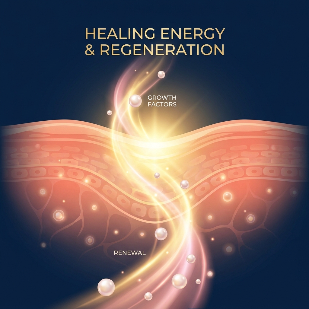
PRP Injection Inflammation Response: The 3-Phase Healing Cascade That Proves Soreness Is the Treatment Working
Introduction: Why Post-PRP Soreness Is Not a Problem — It’s Proof
The morning after a PRP injection, many patients wake up experiencing more discomfort than they felt before the procedure. The knee throbs. The shoulder aches. The immediate thought is often the same: something must have gone wrong. This reaction is understandable, yet it reflects a fundamental misunderstanding of how platelet-rich plasma therapy actually works.
The PRP injection inflammation response is not a side effect to manage or a complication to fear. It is the therapeutic mechanism itself. When patients experience post-injection soreness, swelling, and warmth, they are witnessing their body’s healing cascade activating exactly as intended.
Most clinic content explains what to expect after PRP treatment, listing symptoms like pain and swelling without ever explaining the biological reason these symptoms occur. This article fills that gap by detailing the three-phase healing cascade that transforms initial inflammation into lasting tissue repair. The key biological players in this process include macrophages, growth factors, and cytokines, all working in coordinated sequence.
This response is well-documented in peer-reviewed literature and supported by leading institutions including Johns Hopkins Medicine and Cleveland Clinic. Understanding the science behind post-PRP inflammation empowers patients to support their recovery rather than work against it.
What PRP Actually Is and Why Inflammation Is Built Into the Design
PRP is produced by centrifuging a patient’s own blood to concentrate platelets to two to nine times their normal baseline level. This concentrated platelet solution is then injected directly into the injured area to stimulate tissue repair.
The therapeutic power of PRP lies in what platelets contain. Each platelet carries alpha-granules packed with growth factors, including PDGF (Platelet-Derived Growth Factor), TGF-β (Transforming Growth Factor-beta), VEGF (Vascular Endothelial Growth Factor), EGF (Epidermal Growth Factor), FGF (Fibroblast Growth Factor), and IGF-1 (Insulin-like Growth Factor 1). When injected, these growth factors are released in supraphysiological concentrations at the treatment site.
The critical concept to understand is this: PRP is intentionally designed to trigger the body’s natural inflammatory response as the first step in a coordinated healing cascade. This stands in direct contrast to corticosteroid injections, which suppress inflammation. PRP and corticosteroids operate through fundamentally opposite mechanisms.
The soreness patients feel after PRP is not the treatment failing. It is the treatment activating.
The 3-Phase Healing Cascade: A Timeline of What Is Happening Inside the Body
The healing process following PRP injection follows a predictable three-phase model. Each phase is sequential and interdependent. Suppressing or skipping Phase 1 compromises the outcomes of Phases 2 and 3. This framework is supported by peer-reviewed literature, including research published in PMC documenting how PRP therapy supports the inflammation, proliferation, and remodeling phases of tissue healing.
Phase 1: Inflammation (Days 1 to 7): The Storm That Starts the Repair
During the first week following injection, patients typically experience increased pain, swelling, warmth, and stiffness. These symptoms generally peak within 48 to 72 hours before gradually subsiding.
The biology behind this phase is straightforward. When PRP is injected, platelet alpha-granules release their growth factor payload at the injection site, triggering a localized inflammatory cascade. This cascade recruits M1 macrophages to the area. These pro-inflammatory immune cells are the body’s first responders, performing phagocytosis (clearing cellular debris) and apoptosis of damaged tissue.
PDGF plays a particularly important role during this phase as a potent chemoattractant, drawing macrophages and fibroblasts to the injury site. The pain and swelling patients experience are signals of active immune recruitment. The body is mobilizing its repair resources.
For most patients, this phase resolves within the first week.
Phase 2: Proliferation (Weeks 2 to 6): New Tissue Takes Shape
As the acute inflammatory phase subsides, patients typically notice a gradual reduction in pain, early return of function, and a sense that the tissue is beginning to feel different.
The defining event of this phase is the M1-to-M2 macrophage transition. M2 macrophages are anti-inflammatory cells that replace their M1 counterparts and shift the biological environment from destruction to construction.
TGF-β becomes particularly active during this phase, attracting fibroblasts, stimulating collagen synthesis, and promoting angiogenesis (new blood vessel formation). VEGF drives neovascularization, creating new blood vessels that deliver oxygen and nutrients to the repair site. PRP also recruits local stem cells and fibroblasts to the damaged area.
During this phase, PRP begins to downregulate inflammatory cytokines such as IL-1β, IL-6, and TNF-α. The environment transitions from pro-inflammatory to anti-inflammatory. This is why patients often notice meaningful improvement in pain and mobility between weeks two and six.
Phase 3: Remodeling (Weeks 6 and Beyond): Functional Restoration
The remodeling phase brings significant functional improvement, reduced pain, and return to activity. Full recovery, however, typically requires three to six months.
During remodeling, newly formed collagen fibers are reorganized and strengthened along lines of mechanical stress. EGF and FGF continue to support cell proliferation and matrix maturation. Tendon injuries typically require longer remodeling timelines than joint or soft tissue injuries.
The quality of Phase 3 outcomes depends directly on the integrity of Phase 1. Disrupting early inflammation compromises long-term repair. Patients should expect six to eight weeks to assess initial response and three to six months for full recovery, a timeline detailed in our PRP therapy recovery timeline guide.
The Macrophage Switch: The Biological Turning Point Most Patients Never Hear About
The M1-to-M2 macrophage polarization represents the most compelling and underexplained mechanism in PRP healing.
M1 macrophages function as first responders. They clear debris and signal the immune system, generating the initial soreness and swelling patients experience. M2 macrophages serve as the construction crew, coordinating tissue repair, promoting fibroblast proliferation, and resolving inflammation.
PRP’s growth factor cocktail, particularly PDGF and TGF-β, orchestrates this macrophage polarization shift. Research published in PMC examines how leukocyte content and macrophage function influence the healing cascade.
The clinical implication is clear: the M1 phase (soreness) is not the enemy. It is the prerequisite for the M2 phase (healing). A useful analogy frames the M1 phase as demolition before construction, while the M2 phase represents the building process itself.
The Growth Factor Cascade: What Each Molecule Does and When
Understanding the specific growth factors in PRP provides insight into how healing unfolds:
PDGF (Platelet-Derived Growth Factor): A potent chemoattractant for macrophages and fibroblasts that stimulates collagen synthesis and enhances TGF-β secretion. Active primarily in Phase 1 and early Phase 2.
TGF-β (Transforming Growth Factor-beta): Attracts fibroblasts, stimulates collagen synthesis, promotes angiogenesis, and helps downregulate excessive inflammation. Bridges Phase 1 and Phase 2.
VEGF (Vascular Endothelial Growth Factor): Drives new blood vessel formation, delivering nutrients to the repair site. Critical in Phase 2 and also produced by leukocytes in leukocyte-rich PRP.
EGF (Epidermal Growth Factor): Stimulates cell proliferation and differentiation, supporting tissue maturation in Phases 2 and 3.
FGF (Fibroblast Growth Factor): Promotes fibroblast proliferation and angiogenesis across Phases 2 and 3.
IGF-1 (Insulin-like Growth Factor 1): Supports cell survival, protein synthesis, and tissue growth throughout the healing cascade.
Growth factor levels degrade over time after preparation, making prompt administration following centrifugation clinically important. For a detailed walkthrough of how PRP is prepared and administered, see our PRP injection procedure step-by-step overview.
LR-PRP vs. LP-PRP: How Leukocyte Content Shapes the Inflammatory Response
A fundamental distinction exists between Leukocyte-Rich PRP (LR-PRP), which contains white blood cells, and Leukocyte-Poor PRP (LP-PRP), which has most leukocytes removed. Leukocyte content is one of the primary variables determining the intensity and duration of the post-injection inflammatory response.
LR-PRP triggers a stronger initial inflammatory response. Leukocytes contribute to immune defense, cell chemotaxis, and VEGF production, making this formulation potentially more appropriate for certain tendon injuries or infection-prone environments.
LP-PRP is associated with a more anti-inflammatory cytokine profile and may produce less post-injection soreness. It may be better tolerated in cartilage environments where catabolic activity is a concern, as in vitro studies show high leukocyte content can upregulate IL-1 and TNF-α.
A landmark double-blind randomized trial of 192 knee osteoarthritis patients found that both LR-PRP and LP-PRP produced similar clinical improvement at 12 months. A 2025 BMC Musculoskeletal Disorders study in rabbits also found similar therapeutic effects in early-stage knee osteoarthritis, with LP-PRP noted for its safety profile.
If post-PRP soreness is more intense, the provider may have used LR-PRP. This represents a clinical decision, not an error. Treatment selection should be personalized based on injury type, location, and patient factors. This personalized approach is central to how Unicorn Bioscience designs personalized regenerative medicine protocols.
Why NSAIDs Are Contraindicated After PRP and What to Use Instead
One of the most common questions patients have after PRP is also one of the least clearly answered: what can be taken for pain?
NSAIDs such as ibuprofen, naproxen, and aspirin work by inhibiting COX enzymes and blocking prostaglandin synthesis. This is the same biochemical pathway that drives the inflammatory response PRP is designed to trigger.
Taking an NSAID after PRP is pharmacologically equivalent to applying a brake while the treatment is trying to accelerate. It directly undermines the therapeutic mechanism.
Appropriate pain management alternatives include:
- Acetaminophen (Tylenol) for pain relief
- Ice for localized comfort (with provider guidance)
- Rest and activity modification
Patients should discuss their full medication list with their provider before treatment. NSAID avoidance is typically recommended for a period before and after injection; patients should follow their specific provider’s protocol.
Normal Inflammation vs. Warning Signs: How to Read the Body’s Signals
While post-PRP inflammation is expected and therapeutic, patients need a clear framework to distinguish normal healing from genuine complications.
Normal inflammatory response:
- Increased pain, swelling, warmth, and stiffness peaking at 48 to 72 hours
- Gradual improvement within 7 days
- Manageable with acetaminophen and rest
Signs that warrant contacting a provider:
- Pain that continues to worsen beyond 10 to 14 days without any improvement
- Fever or systemic symptoms
- Redness spreading beyond the injection site
- Signs consistent with infection
Pre-existing inflammation at the injection site, such as severe arthritis or advanced tendon degeneration, can intensify and prolong the post-PRP inflammatory response. This is expected and not a sign of treatment failure.
The presence of inflammation is reassuring. The absence of any response, or a response that escalates without plateau, warrants clinical attention.
Factors That Influence the Intensity of the PRP Inflammation Response
Not all patients experience the same intensity or duration of post-PRP inflammation. This variability is clinically meaningful:
- PRP formulation: LR-PRP vs. LP-PRP is one of the strongest predictors of inflammatory intensity.
- Platelet concentration: Higher concentrations release more growth factors and may trigger a more robust initial response.
- Activation status: Activated PRP releases growth factors immediately; non-activated PRP releases them more gradually.
- Injury severity: Patients with more advanced degeneration or chronic inflammation typically experience a more pronounced response.
- Patient age and immune status: Immune function changes affect macrophage recruitment and the M1-to-M2 transition timeline.
- Injection site: Tendon injections often produce more intense and prolonged soreness than intra-articular injections. Conditions such as Achilles tendonitis are a common example where this extended response is expected.
Emerging research suggests individual genetic variations in TGF-β, PDGF, or VEGF receptor genes may influence PRP responsiveness. Personalized treatment protocols, such as those used at Unicorn Bioscience, account for these variables to optimize the inflammatory response for each patient.
The Serotonin Connection: An Emerging Dimension of PRP’s Immune Modulation
Platelet-derived serotonin (5-HT) represents an underappreciated component of PRP’s biological activity. Platelets are the primary storage and release vehicle for serotonin in the bloodstream.
Serotonin acts as a regulator of both innate and adaptive immune responses, influencing macrophage behavior and cytokine production. It may play a role in modulating the transition from the M1 to M2 macrophage phase, potentially influencing how quickly the inflammatory response resolves.
This is an emerging research area not yet fully characterized in clinical PRP literature. It does, however, demonstrate that PRP’s mechanism is more complex and multifaceted than a simple growth factor injection.
What This Means for Recovery: Supporting the Healing Cascade, Not Fighting It
Practical guidance helps patients work with the healing cascade rather than against it:
Activity modification: Patients should avoid high-impact loading of the treated area during Phase 1 and follow provider guidance for a gradual return to activity during Phase 2.
Nutrition: Adequate protein intake supports collagen synthesis during Phases 2 and 3. Hydration supports overall cellular function.
Sleep: Growth hormone secretion during sleep supports tissue repair.
Avoid anti-inflammatory medications and supplements (including high-dose fish oil, turmeric supplements, and NSAIDs) during the critical inflammatory window unless specifically cleared by a provider.
Follow-up appointments: The six to eight week mark is typically when providers assess initial treatment response. Patients should not judge outcomes before this point.
Full recovery takes three to six months. Tendon injuries may require longer timelines. Some patients benefit from a series of injections rather than a single treatment. The intensity of the early inflammatory response is not a reliable predictor of final outcomes.
Conclusion: Inflammation Is the Treatment
Post-PRP inflammation is not a complication to suppress. It is the biological mechanism through which healing occurs.
The three-phase cascade unfolds predictably: Phase 1 (inflammation, Days 1 to 7), Phase 2 (proliferation, Weeks 2 to 6), and Phase 3 (remodeling, Weeks 6 and beyond). The key biological players include the M1-to-M2 macrophage transition, PDGF, TGF-β, VEGF, EGF, and the role of leukocyte content in shaping the response.
Blocking inflammation blocks the treatment. Acetaminophen and provider-guided supportive care are the appropriate alternatives to NSAIDs.
The LR-PRP vs. LP-PRP distinction reflects the nuance of personalized medicine. There is no single best formulation, only the right formulation for the right patient.
Patients who understand the biology of their treatment are better equipped to support it, set realistic expectations, and communicate meaningfully with their care team. Unicorn Bioscience’s approach centers on personalized treatment protocols, precision-guided injection delivery, and comprehensive patient education to optimize outcomes.
Ready to Understand Your Treatment, Not Just Undergo It?
Patients who seek this level of biological understanding deserve a provider who delivers it. Unicorn Bioscience offers consultations in person across eight locations in Texas, Florida, and New York, or virtually for those who prefer remote access.
Unicorn Bioscience’s personalized treatment protocols account for individual factors including inflammation levels, age, injury type, and health goals. PRP is one of several regenerative options available, including stem cell therapy, BMAC, exosomes, and hyaluronic acid, depending on the patient’s condition.
To schedule a consultation, call (737) 347-0446 or visit unicornbioscience.com.
Schedule Your Consultation Today!
