Ache Pain Behind Knee: The 8-Cause Posterior Fossa Map That Tells You When to Wait and When to Act
Ache Pain Behind Knee: The 8-Cause Posterior Fossa Map That Tells You When to Wait and When to Act
Introduction: That Nagging Ache Behind Your Knee and Why It Matters Which Kind It Is
The scenario is all too familiar. A dull, persistent ache settles into the back of the knee after a long walk, a morning run, or simply sitting at a desk for hours. The discomfort lingers, prompting questions: Is this something serious? Should a doctor be called? Or will it simply go away on its own?
Knee pain affects approximately 25% of adults, and its prevalence has increased almost 65% over the past two decades. According to the American Academy of Family Physicians, knee complaints account for nearly 4 million primary care visits annually in the United States. Posterior knee pain, the aching or achy pain behind the knee, represents a distinct and often misunderstood subset of these cases.
The popliteal fossa serves as the anatomical “map” for understanding this type of discomfort. This diamond-shaped region at the back of the knee houses an intricate network of tendons, muscles, nerves, blood vessels, and bursae. Any of these structures can generate the nagging ache pain behind the knee that brings patients to their physicians.
This article organizes the eight major causes of posterior knee aching into a triage-first framework. Rather than simply listing conditions, it addresses not just what the cause might be, but what should be done about it today. The framework divides causes into three urgency tiers: watch-and-wait, seek care soon, and urgent/emergency. By understanding which tier applies to their symptoms, readers can immediately orient themselves toward the appropriate course of action.
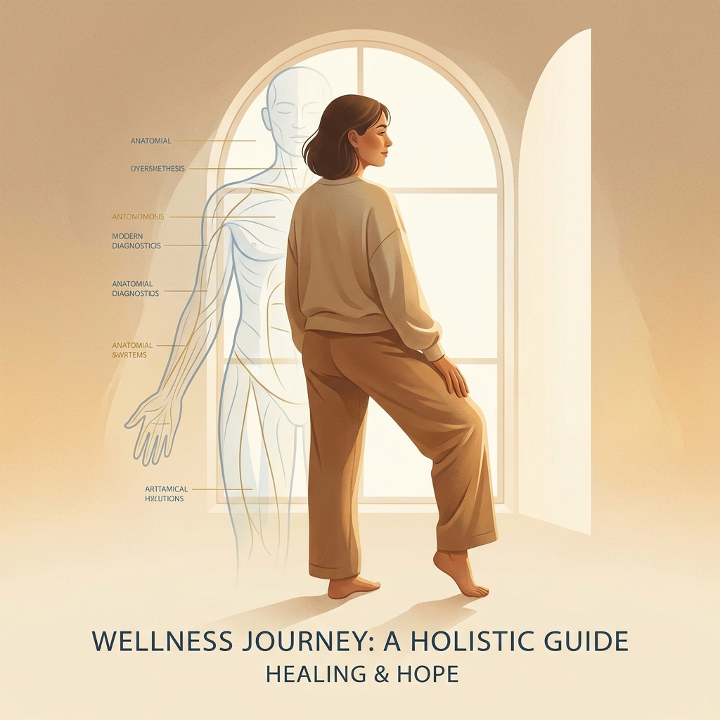
Understanding the Posterior Knee: A Quick Anatomy Orientation
The popliteal fossa contains a remarkable concentration of anatomical structures within a relatively small space. The hamstring tendons (biceps femoris, semimembranosus, and semitendinosus) form the upper boundaries. The gastrocnemius muscle heads create the lower borders. Deep within this space lie the popliteus muscle, the posterior joint capsule, the posterior horns of the menisci, and the posterior cruciate ligament.
The popliteal artery and vein traverse this region, along with the tibial nerve and the popliteal bursa. This convergence of soft tissue, vascular, and neural elements explains why so many different conditions can produce a similar aching sensation in this location.
A 2025 research finding underscores the complexity of posterior knee pain diagnosis. In 72.5% of cases involving posterior knee pain during straight leg raise tests, neural structures were primarily responsible for the discomfort rather than muscular tension. This often-overlooked finding demonstrates why self-diagnosis based on location alone is insufficient.
The key to distinguishing between causes lies in three axes: the character of the ache (dull versus throbbing versus sharp), the context (rest versus movement versus specific positions), and associated symptoms. These factors guide the diagnostic process that follows.
The Posterior Knee Ache Triage System: Three Urgency Tiers
Understanding the urgency of posterior knee pain requires a systematic framework. This triage system organizes all eight causes by the appropriate level of response.
Tier 1: Watch and Wait encompasses conditions that typically respond to self-management at home over one to two weeks with conservative care. This tier includes Baker’s cyst, hamstring tendinopathy, gastrocnemius strain, and popliteus tendinopathy.
Tier 2: Seek Care Soon includes conditions requiring a medical appointment within days to one to two weeks, with imaging likely needed. This tier covers posterior meniscal tears, PCL injuries, and osteoarthritis flares.
Tier 3: Urgent/Emergency demands same-day or emergency care. Deep vein thrombosis (DVT) and popliteal vein thrombosis fall into this category.
The rationale for this system is straightforward. Most causes of aching pain behind the knee are benign and self-limiting. However, one cause, DVT, can be life-threatening if missed. The symptom overlap between DVT and a ruptured Baker’s cyst makes this distinction critical.
Red Flag Checklist: Any of the following symptoms warrants urgent evaluation: sudden severe swelling, warmth and redness, inability to bear weight, visible deformity, a popping sound at the time of injury, or pain worsening progressively over days.
Tier 1: Watch and Wait: Four Causes of Posterior Knee Aching That Usually Resolve With Conservative Care
These four causes represent the most common sources of ache pain behind the knee in adults. They typically respond well to rest, activity modification, ice, NSAIDs, and physical therapy over one to four weeks. Importantly, “watch and wait” does not mean “ignore.” It means active self-management with a defined timeline and clear criteria for escalating to professional care.
Cause 1: Baker’s Cyst (Popliteal Cyst): The Most Common Culprit
A Baker’s cyst is a fluid-filled sac that forms in the popliteal fossa. Most common in adults aged 35 to 70, it is strongly associated with underlying osteoarthritis and meniscal tears. Research indicates that popliteal cysts have been found in 4.7% to 37% of adults with asymptomatic knees.
The symptom profile includes dull, achy pressure or fullness behind the knee that worsens with prolonged standing, bending, or after activity. Patients may feel a soft lump and experience stiffness after sitting.
The critical distinction involves rupture risk. A ruptured Baker’s cyst can closely mimic DVT, presenting with swelling, calf pain, and warmth. Duplex ultrasonography serves as the first-line diagnostic tool to exclude DVT.
Action: If the ache is mild without sudden worsening of swelling or warmth, conservative management (ice, elevation, NSAIDs, and addressing the underlying cause) is appropriate for one to two weeks. If swelling suddenly increases or the calf becomes red and warm, escalate immediately to Tier 3 urgent care. Treating the underlying cause remains essential; the cyst is a symptom, not the root problem. For a deeper look at back of knee joint pain and how it relates to conditions like Baker’s cyst, additional guidance is available.
Cause 2: Hamstring Tendinopathy (Distal): The Runner’s Ache
Distal hamstring tendinopathy involves degeneration or irritation of the distal hamstring tendons, most commonly the semimembranosus or biceps femoris, where they attach near the back of the knee.
Patients experience localized achy pain at the back of the knee that worsens with bending or straightening the leg, running, cycling, or prolonged sitting. Tenderness on palpation of the tendon insertion and stiffness after rest that loosens with gentle movement are characteristic.
This condition commonly affects runners, cyclists, and athletes performing repetitive knee flexion. It also occurs in sedentary adults who suddenly increase activity.
Action: Relative rest, ice, and NSAIDs address the acute phase. Progressive loading exercises form the cornerstone of recovery, with return-to-activity timelines typically spanning four to eight weeks. If pain persists beyond four weeks of consistent conservative care, imaging (ultrasound or MRI) is warranted to rule out partial tears.
Cause 3: Gastrocnemius Muscle Strain: When the Calf Aches Upward
A strain or tightness of the gastrocnemius muscle, which originates from the posterior femoral condyles just above the knee joint, can radiate an aching pain upward into the popliteal fossa.
The symptom profile includes aching pain behind the knee and upper calf, especially after running, jumping, or prolonged walking. Pain may worsen with ankle dorsiflexion (pulling toes up) and is often accompanied by calf tightness.
The key distinction from DVT: gastrocnemius strain pain is typically activity-related and improves with rest. DVT pain is often constant and associated with swelling, warmth, and redness.
Action: RICE (rest, ice, compression, elevation) and gentle stretching once the acute phase resolves typically lead to resolution within one to three weeks. If swelling is disproportionate to the injury or accompanied by warmth and redness, rule out DVT urgently.
Cause 4: Popliteus Tendinopathy: The Downhill Runner’s Ache
Popliteus tendinopathy involves irritation or degeneration of the popliteus tendon, a small but important stabilizing muscle that runs diagonally across the back of the knee.
The symptom profile features aching pain localized to the outer-back corner of the knee (posterolateral), characteristically worsening with downhill running or walking, prolonged standing, or knee hyperextension.
This condition is frequently misdiagnosed as lateral meniscal pathology or IT band syndrome. The posterolateral location and downhill aggravation are the key distinguishing features.
Action: Activity modification (avoiding downhill running temporarily), eccentric strengthening, and addressing biomechanical contributors such as overpronation and hip weakness typically resolve symptoms in four to eight weeks. Persistent pain beyond six weeks or suspected posterolateral corner injury requires imaging and specialist evaluation.
Tier 2: Seek Care Soon: Three Causes That Need Professional Evaluation and Likely Imaging
These three causes involve structural damage to cartilage, ligaments, or joints that cannot be adequately assessed or managed without professional evaluation, imaging, and a tailored treatment plan. The aching pain pattern alone may not distinguish these from Tier 1 causes. The key differentiators are mechanism of injury, associated symptoms (locking, giving way, morning stiffness), and failure to improve with one to two weeks of conservative care.
Cause 5: Posterior Horn Meniscal Tear: The Deep, Catching Ache
A tear in the posterior horn of the medial or lateral meniscus affects the cartilage “shock absorbers” of the knee. Posterior horn tears represent the most common location for meniscal tears.
Patients experience deep, aching pain behind the knee (often medial or lateral) along with catching, clicking, or locking sensations with bending or twisting. Pain with squatting, kneeling, or climbing stairs is common, and swelling may occur after activity.
MRI is the gold standard for soft tissue posterior knee pathology. According to JAMA research, conservative management (exercise therapy for four to six weeks) is appropriate for most degenerative meniscal tears. Surgery is not the automatic first step.
Notably, imaging findings do not always correlate with symptoms. A tear visible on MRI may not be the source of pain, which is why clinical evaluation matters.
For patients who have not responded to conservative care, regenerative medicine options such as PRP injections and cellular therapies are emerging as non-surgical alternatives. Unicorn Bioscience offers these treatments as part of a comprehensive approach to meniscus repair with stem cell therapy and meniscal degeneration.
Cause 6: Posterior Cruciate Ligament (PCL) Injury: The Often-Missed Ligament Ache
The PCL serves as the primary restraint against posterior tibial translation. While less common than ACL injuries, PCL injuries cause significant posterior knee pain and instability.
The symptom profile includes aching or throbbing pain deep in the back of the knee, a feeling of instability or “wobbliness,” swelling within hours of injury, and pain worsening with stairs, kneeling, or pivoting.
PCL injuries typically occur from direct impact to the front of the knee (such as a dashboard injury in car accidents), falling onto a bent knee, or hyperextension. MRI has demonstrated 90.4% sensitivity for detecting PCL damage and remains the gold standard for diagnosis.
Action: Isolated PCL injuries are generally treated conservatively with bracing, physical therapy, and progressive strengthening. Surgical reconstruction is reserved for complete tears with instability or multi-ligament injuries. If the knee is significantly swollen, unable to bear weight, or there was a high-energy mechanism, seek care urgently.
Cause 7: Knee Osteoarthritis: The Chronic, Activity-Related Ache in Adults 45 and Older
Knee osteoarthritis involves progressive degeneration of articular cartilage in the knee joint. According to JAMA, OA is the most likely diagnosis of knee pain in patients aged 45 or older presenting with activity-related pain and less than 30 minutes of morning stiffness.
While OA pain is often diffuse, it frequently manifests as an aching pain behind the knee due to posterior joint capsule involvement, posterior osteophytes, and secondary Baker’s cyst formation.
The numbers are striking. Knee OA affects an estimated 654 million people worldwide aged 40 and older. In 2021, it accounted for 374.74 million prevalence cases globally. The lifetime risk of symptomatic knee OA is approximately 45%, and nearly 37% of adults 65 and older report knee pain.
Risk factors include age, female sex (prevalence ratio of approximately 1.69 compared to males), obesity or elevated BMI (a modifiable risk factor often underrepresented in patient education), and prior joint injury. Understanding what causes knee pain in females can help contextualize these risk differences.
First-line treatment is conservative: exercise therapy, weight management, NSAIDs, and activity modification. When conservative care fails, the treatment ladder expands to include injections, regenerative therapies, and, as a last resort, surgery.
Unicorn Bioscience offers a comprehensive regenerative medicine pathway for OA patients, including PRP, stem cell therapy (BMAC), hyaluronic acid injections, and exosome therapy. The clinic reports that more than 90% of their stem cell patients have not gone on to knee replacement surgery. This aligns with broader research suggesting that up to 80% of patients told they need total knee replacement may not actually require surgery.
Tier 3: Urgent/Emergency: The One Cause of Posterior Knee Aching That Cannot Be Missed
One cause of ache pain behind the knee can be life-threatening if not treated promptly. DVT can look deceptively similar to a Baker’s cyst or muscle strain, which is why it deserves explicit discussion.
Cause 8: Deep Vein Thrombosis (DVT) / Popliteal Vein Thrombosis: The Emergency That Must Be Ruled Out
A blood clot in the popliteal vein or other deep veins of the leg can cause aching, throbbing pain behind the knee and in the calf.
The symptom profile includes throbbing or aching pain behind the knee and calf, swelling (often asymmetric, with one leg more swollen than the other), warmth and redness over the affected area, and pain that does not clearly improve with rest.
The silent danger: DVT may not cause symptoms in approximately 30 to 40% of cases, making clinical vigilance essential.
The Baker’s cyst confusion presents a significant diagnostic challenge. A ruptured Baker’s cyst can closely mimic DVT, as both cause sudden calf swelling, warmth, and pain. Duplex ultrasonography is the first-line diagnostic tool to exclude DVT and should be performed before assuming a cyst has ruptured.
Risk factors include prolonged immobility (long flights, bed rest), recent surgery, pregnancy, oral contraceptives, cancer, personal or family history of clotting disorders, and obesity.
Action: Seek same-day emergency care if experiencing sudden severe swelling in one leg, warmth and redness behind the knee or calf, constant pain not relieved by rest, or any combination of these symptoms. Do not wait. Untreated DVT can lead to pulmonary embolism, a potentially fatal condition.
The Posterior Knee Ache Symptom-to-Cause Quick Reference Map
| Symptom Pattern | Likely Cause | Tier |
|---|---|---|
| Dull, achy pressure behind the knee with feeling of fullness or soft lump | Baker’s cyst | 1 (watch and wait unless sudden worsening) |
| Localized aching at the back of the knee worsening with running or bending | Hamstring tendinopathy | 1 (conservative care) |
| Aching behind the knee and upper calf after running or jumping | Gastrocnemius strain | 1 (RICE protocol) |
| Aching at the outer-back corner of the knee, worse downhill | Popliteus tendinopathy | 1 (activity modification plus rehab) |
| Deep aching behind the knee with catching, clicking, or locking | Posterior meniscal tear | 2 (seek care within 1 to 2 weeks) |
| Aching deep in the knee after a direct blow or fall, with instability | PCL injury | 2 (seek care promptly) |
| Dull, activity-related aching in adults 45 and older, worse with stairs, morning stiffness under 30 minutes | Osteoarthritis | 2 (seek care for management plan) |
| Throbbing ache behind the knee with swelling, warmth, redness, or asymmetric leg swelling | DVT | 3 (seek emergency care immediately) |
This map serves as a starting point, not a substitute for professional diagnosis. Given the 2025 finding that neural structures are responsible for posterior knee pain in 72.5% of straight leg raise cases, the source of pain is not always where it feels. Noting specific symptom patterns before appointments helps providers narrow the diagnosis efficiently.
First-Line Treatment for Posterior Knee Aching: What Works Before Seeing a Doctor
For Tier 1 causes and as a bridge while awaiting a Tier 2 appointment, evidence-based self-care can meaningfully reduce aching pain behind the knee.
RICE Protocol: Rest, ice (15 to 20 minutes, three to four times daily), compression, and elevation remain appropriate for acute soft tissue causes.
NSAIDs: Ibuprofen and naproxen reduce inflammation and pain for most soft tissue and OA-related causes. Contraindications include kidney disease, GI issues, and blood thinner use. These medications should not mask symptoms of DVT.
Activity Modification: Identifying and temporarily reducing the specific activity that aggravates the ache (running, squatting, prolonged sitting) is essential. Complete rest is rarely necessary and often counterproductive.
Gentle Movement and Stretching: For hamstring tendinopathy and gastrocnemius strain, gentle range-of-motion exercises maintain mobility without aggravating the tendon.
Heat Versus Ice: Ice is preferred in the first 48 to 72 hours for acute strains. Heat may help chronic stiffness (such as OA-related morning stiffness) after the acute phase.
Weight Management: For OA-related posterior knee aching, even modest weight loss significantly reduces joint load.
Timeline: If symptoms do not improve meaningfully within one to two weeks of consistent conservative care, a professional evaluation should be scheduled.
What Not to Do: Do not ignore sudden swelling, warmth, or redness. Do not continue high-impact activity through worsening pain. Do not self-diagnose and delay care if red-flag symptoms are present.
When Conservative Care Is Not Enough: The Non-Surgical Treatment Pathway
For Tier 2 causes that do not resolve with conservative care, a structured non-surgical treatment pathway exists before surgery should be considered.
Physical Therapy and Progressive Loading: This remains the cornerstone of non-surgical management for tendinopathy, meniscal degeneration, and OA. Individualized programs address strength, flexibility, and biomechanics.
Corticosteroid Injections: These provide short-term pain relief for OA and bursitis but are not a long-term solution and may have cartilage-damaging effects with repeated use.
Hyaluronic Acid Injections: Viscosupplementation restores joint lubrication in OA and is available at regenerative medicine clinics including Unicorn Bioscience. Patients often ask about hyaluronic acid vs stem cells when weighing their options.
Regenerative Medicine Options: The next step in the non-surgical ladder includes PRP (concentrated platelets from the patient’s own blood), stem cell therapy/BMAC (using the patient’s own bone marrow cells), and exosome therapy (extracellular vesicles that facilitate cellular communication). Understanding the difference between PRP and stem cell therapy can help patients make informed decisions. Currently, 224 clinical trials globally are investigating stem cell therapies for osteoarthritis, and a major Phase III clinical trial funded with $140 million was announced in January 2026.
Genicular Nerve Radiofrequency Ablation: Evidence from 2025 and 2026 supports this as an effective non-surgical option for chronic knee OA pain when other measures fail.
As of 2026, the FDA has not approved stem cell, PRP, or exosome products specifically for orthopedic conditions. However, substantial clinical evidence supports safety and efficacy when administered by qualified providers within FDA regulatory frameworks.
How Unicorn Bioscience Fits Into the Posterior Knee Pain Journey
For patients with Tier 2 causes (OA, meniscal degeneration, tendinopathy) who have not responded to conservative care and want to explore non-surgical options, Unicorn Bioscience offers a comprehensive regenerative medicine approach.
The clinic provides a multi-modal regenerative medicine treatment approach including PRP, stem cell therapy (BMAC), exosome therapy, hyaluronic acid injections, and peptide therapy. Each treatment plan is customized to the individual patient’s inflammation levels, age, injury type, and health goals.
All injections are administered using precision-guided technology, including ultrasound-guided injection for joint pain and X-ray guidance, ensuring accurate delivery to posterior knee structures. Qualified candidates can receive same-day orthopedic injection treatment, reducing the time between diagnosis and treatment initiation.
With eight locations across Texas, Florida, and New York, plus virtual consultation availability, access is practical for a broad patient population.
When to See a Doctor: A Clear Decision Framework
See a doctor today or go to the ER if: Sudden severe swelling occurs in one or both legs; warmth and redness appear behind the knee or calf; constant throbbing pain is not relieved by rest; weight-bearing is impossible; visible deformity is present; or a popping sound occurred at the time of injury.
Schedule an appointment within one to two weeks if: Aching pain behind the knee has not improved after one to two weeks of consistent self-care; pain is associated with catching, locking, or giving way; pain followed a direct blow or fall; or the patient is 45 or older with new-onset activity-related posterior knee aching.
Continue self-managing (watch and wait) if: Mild to moderate achy pain behind the knee is clearly activity-related, improving with rest and ice, without swelling, warmth, redness, or instability, and no red-flag symptoms are present.
This framework is a guide, not a substitute for professional medical evaluation. When in doubt, err on the side of seeking care. If uncertainty exists about whether symptoms represent a ruptured cyst or a blood clot, treat it as a potential DVT and seek same-day evaluation.
Conclusion: A Posterior Knee Ache Has a Pattern and a Path Forward
Eight causes of ache pain behind the knee, organized into three urgency tiers, each with a specific action recommendation: this framework empowers patients to make informed decisions about their care.
Most causes of aching pain behind the knee are benign and respond well to conservative care. However, accurate identification of the cause is essential to choosing the right treatment and avoiding the one cause (DVT) that requires emergency attention.
The posterior knee is a dense anatomical region where soft tissue, vascular, and neural structures converge. The 2025 finding that neural structures drive posterior knee pain in 72.5% of cases serves as a reminder that professional evaluation adds value that self-diagnosis cannot replicate.
For the majority of patients with Tier 2 causes, surgery is not the inevitable outcome. Physical therapy, regenerative medicine, and minimally invasive interventions offer meaningful alternatives for those experiencing persistent achy pain behind the knee. Patients exploring knee osteoarthritis cellular therapy will find a growing body of evidence supporting these approaches.
Understanding the specific ache pattern, including its character, context, and associated symptoms, puts patients in a stronger position to have informed conversations with their providers and advocate for the right care.
Ready to Stop Guessing About Posterior Knee Pain?
If aching pain behind the knee has persisted beyond one to two weeks of self-care, or if surgery has been presented as the only option, Unicorn Bioscience offers a non-surgical alternative worth exploring.
Virtual and in-person consultations are available at eight locations across Texas, Florida, and New York, with same-day treatment available for qualified candidates. Treatment plans are developed based on individual factors including inflammation levels, age, injury type, current medications, and personal health goals.
For more information, call (737) 347-0446 or visit unicornbioscience.com.
Patients can use the symptom-to-cause map from this article as a starting point for the consultation conversation. Determining whether regenerative medicine is appropriate for a specific situation begins with a single step.
Schedule Your Consultation Today!