
Wrist Injury Regenerative Treatment: The Anatomy-First Injection Framework That Matches Every Condition to the Right Biologic
Wrist Injury Regenerative Treatment: The Anatomy-First Injection Framework That Matches Every Condition to the Right Biologic
Introduction: Why the Wrist Demands a Different Regenerative Approach
Emergency department visits for sports-related soft tissue injuries increased by 22% in 2025, including those affecting the wrist. The patient population seeking treatment now extends far beyond competitive athletes to include office workers, gamers, and remote employees dealing with repetitive strain from keyboards and handheld devices.
Most patients researching wrist injury regenerative treatment encounter generic descriptions of PRP or stem cells without understanding why the wrist’s anatomy makes protocol selection and injection precision uniquely critical. The wrist is not simply a smaller version of the knee or hip. Its complexity demands a fundamentally different approach.
The wrist’s anatomical complexity, including eight carpal bones, the triangular fibrocartilage complex (TFCC), the carpal tunnel, scapholunate ligaments, and avascular zones, is not merely background information. It serves as the primary driver of which biologic is chosen, how it is delivered, and why outcomes differ dramatically between conditions.
Conventional treatments such as splints, corticosteroid injections, and NSAIDs provide temporary symptom relief but do not promote actual tissue repair at the biological level. Regenerative treatments like PRP and bone marrow aspirate concentrate (BMAC) are designed to address the underlying pathology rather than mask symptoms.
This article walks through each major wrist condition, explains the anatomical rationale behind the regenerative protocol selected, and outlines what the clinical evidence shows. Readers will gain the information needed to have an informed conversation with a specialist. Unicorn Bioscience applies this anatomy-first, precision-guided approach across all eight clinic locations in Texas, Florida, and New York.
The Anatomical Blueprint: Why the Wrist Is Unlike Any Other Joint
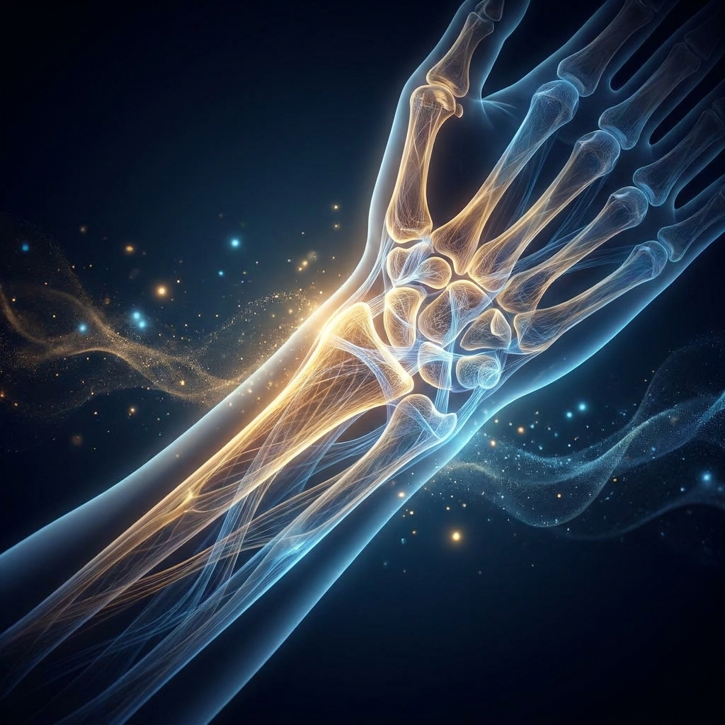
The wrist contains eight small carpal bones arranged in two rows: the scaphoid, lunate, triquetrum, and pisiform in the proximal row, and the trapezium, trapezoid, capitate, and hamate in the distal row. These bones are connected by an intricate web of ligaments and stabilized by the distal radius and ulna.
The TFCC is a multi-layered structure on the ulnar side of the wrist that acts as a load-bearing cushion and stabilizer. Critically, its central zone has inherently poor blood supply, making natural healing difficult and biologic delivery strategy essential. MRI studies have demonstrated TFCC injury in 96.5% of patients following distal radius fractures.
The carpal tunnel is a narrow fibro-osseous channel housing the median nerve and nine flexor tendons. Even minor inflammation within this confined space can compress the nerve and produce the hallmark symptoms of carpal tunnel syndrome: numbness, tingling, and weakness in the thumb, index, and middle fingers.
The scapholunate ligament is the most frequently injured wrist ligament and the primary cause of traumatic carpal instability. Partial tears may be amenable to regenerative repair before progressing to full instability and subsequent arthritis.
Certain wrist structures, including the central TFCC and the proximal scaphoid, have limited or no direct blood supply. This means the body’s natural healing cascade is impaired. Delivering concentrated growth factors or stem cells directly into these avascular zones changes the biological environment in ways the body cannot accomplish through vascular means alone.
The hip or knee offers a relatively large target for injection. The wrist’s densely packed small structures mean that even a few millimeters of injection placement error can result in the biologic missing its target entirely. Different tissues within the wrist, including cartilage, fibrocartilage, ligament, tendon sheath, and nerve, have different regenerative needs. A one-size-fits-all biologic approach is anatomically and biologically inappropriate.
The Two Primary Regenerative Pathways: PRP and Stem Cell Therapy
While multiple biologics exist, including PRP, BMAC, adipose-derived stem cells, exosomes, and hyaluronic acid, the two primary pathways for wrist injuries are PRP and mesenchymal stem cell (MSC) therapy. The choice between them is driven by injury type, tissue involved, and severity.
As of 2026, the FDA has not approved stem cell, PRP, or exosome products specifically for orthopedic conditions. However, substantial clinical evidence supports safety and efficacy when administered by qualified providers within FDA regulatory frameworks.
Platelet-Rich Plasma (PRP): The Growth Factor Delivery System
PRP is derived from the patient’s own blood and centrifuged to concentrate platelets to three to five times their normal level, along with a corresponding increase in growth factor concentrations.
PRP releases growth factors including PDGF, VEGF, TGF-β, IGF-1, and FGF. These factors promote angiogenesis (new blood vessel formation), cellular proliferation, collagen synthesis, and extracellular matrix remodeling.
PRP is particularly suited to avascular or hypovascular wrist structures. By delivering a concentrated bolus of growth factors directly into tissues with poor blood supply, such as the TFCC or tendon sheaths, PRP essentially bypasses the vascular limitation that prevents natural healing.
Clinical evidence shows 70 to 80% of PRP patients report long-term pain relief and improved function for wrist disorders, with up to 95% functionality recovery reported in chronic hand and wrist conditions. PRP is typically the first-line regenerative option for mild-to-moderate wrist injuries, tendinopathies, tenosynovitis, and as an adjunct to surgical or arthroscopic repair.
Stem Cell Therapy (BMAC and Adipose-Derived MSCs): Structural Regeneration
The two primary stem cell sources used in wrist regenerative treatment are bone marrow aspirate concentrate (BMAC) drawn from the iliac crest and adipose-derived stem cells (microfat) harvested via mini-liposuction.
MSCs can differentiate into ligamentous, cartilaginous, or osseous tissue depending on the local biological environment. This provides structural repair potential that PRP alone cannot offer. Beyond direct differentiation, stem cells release signaling molecules that stimulate local cell proliferation, angiogenesis, and extracellular matrix production.
Stem cell therapy is typically reserved for more severe wrist injuries, advanced osteoarthritis, significant ligament damage, or cases where PRP alone has not produced sufficient improvement. The combination of microfat with PRP has shown clinical benefit in treating wrist osteoarthritis, including in patients with advanced Stage 4 OA who had failed conservative treatment.
Stem cell therapy for wrist conditions is a same-day outpatient procedure with minimal downtime. Patients typically experience pain relief beginning at three to six weeks post-injection, with tissue regeneration confirmable on follow-up imaging.
Why Ultrasound Guidance Is Non-Negotiable for Wrist Injections
Ultrasound-guided injections achieve accuracy rates over 90%, compared to as low as 40% accuracy for landmark or palpation-guided (blind) injections. This gap is clinically unacceptable when treating the wrist’s small, densely packed structures.
Ultrasound guidance enables real-time visualization of small joint capsules, individual tendon sheaths, nerve bundles, blood vessels, and ligament attachment points. These structures cannot be reliably targeted by feel alone.
The wrist contains the median nerve in the carpal tunnel, the ulnar nerve and artery in Guyon’s canal, and the radial artery. These structures can be damaged by imprecise injection. Ultrasound allows the clinician to visualize and avoid these structures in real time.
In the knee or hip, a few millimeters of placement error may still result in the biologic reaching the target tissue. In the wrist, the same error can mean the injection misses a two to three millimeter tendon sheath entirely or contacts a nerve.
Ultrasound can visualize tendon movement, identify sub-compartmentalization within tendon sheaths (particularly relevant for De Quervain’s), and confirm needle tip position before injection. Ultrasound is radiation-free, real-time, and cost-effective, making it the preferred guidance modality for wrist regenerative procedures at Unicorn Bioscience.
Condition-Specific Regenerative Pathways: Matching the Biology to the Injury
Each major wrist condition has a distinct anatomical profile, biological environment, and healing challenge. The regenerative protocol must be designed around those specifics rather than applied generically.
TFCC Tears: Delivering Growth Factors to an Avascular Zone
The central zone of the TFCC has no direct blood supply, meaning the body’s natural inflammatory healing cascade is severely impaired. Peripheral zone tears have better healing potential due to proximity to the vascular periphery.
PRP is selected as the primary biologic because its concentrated growth factors can be delivered directly into the avascular zone, creating a pro-healing environment that the body cannot generate on its own through vascular means. A 2024 retrospective cohort study examined PRP as an adjunctive treatment to arthroscopy for TFCC injury, confirming PRP’s role in augmenting healing.
Wrist arthroscopy for TFCC injuries carries an 8.8% complication rate (all neurological per a 2025 prospective analysis), reinforcing the clinical rationale for exploring regenerative options before surgery. Ultrasound guidance is critical because the TFCC is a thin, complex structure; precise needle placement into the correct zone requires real-time imaging.
Carpal Tunnel Syndrome: Reducing Inflammation Around the Median Nerve
CTS affects approximately 1 to 5% of the general population and 7.8% of working populations, making it one of the most common wrist conditions in clinical practice. Prevalence is increasing among tech workers, remote workers, and gamers with repetitive strain patterns.
PRP injected into the carpal tunnel delivers anti-inflammatory cytokines and growth factors that reduce synovial inflammation, promote nerve sheath health, and may support myelin repair. This addresses the biological cause rather than simply decompressing the nerve surgically.
Ultrasound guidance is uniquely critical here because the median nerve runs through the carpal tunnel alongside nine flexor tendons. An imprecisely placed injection risks direct nerve injection or tendon injury. Real-time ultrasound visualization of the nerve and needle tip is essential to place the biologic in the correct perineural or subsynovial position.
Patients with mild-to-moderate CTS (confirmed by nerve conduction studies) are typically the best candidates. Severe CTS with significant motor loss may still require surgical decompression.
De Quervain’s Tenosynovitis: Precision Delivery Into a Sub-Compartmentalized Tendon Sheath
De Quervain’s tenosynovitis involves inflammation of the first dorsal compartment tendon sheath, which houses the abductor pollicis longus and extensor pollicis brevis tendons at the radial side of the wrist.
The first dorsal compartment frequently has sub-compartmentalization, a septum dividing the sheath into two separate tunnels. A single injection placed in one sub-compartment may not reach the other, explaining why blind corticosteroid injections often fail.
A 2024 systematic review and meta-analysis confirmed PRP for De Quervain’s tenosynovitis provides better long-term outcomes than corticosteroid injections, with greater VAS pain reduction and DASH score improvement at six months post-injection. Ultrasound can identify the presence of sub-compartmentalization before injection, allowing the clinician to ensure the needle enters each sub-compartment separately.
Scapholunate Ligament Injuries: Regenerative Support for the Wrist’s Primary Stabilizer
Scapholunate ligament injury is the most frequent cause of traumatic carpal instability. Untreated or inadequately treated partial tears can progress to complete rupture, dorsal intercalated segment instability (DISI), and ultimately post-traumatic wrist arthritis.
The scapholunate ligament is a C-shaped structure with three distinct regions. The dorsal portion is the primary stabilizer and has limited intrinsic healing capacity.
For partial scapholunate tears, PRP can be injected into the ligament and periligamentous tissue to deliver growth factors that stimulate fibroblast proliferation and collagen synthesis. For more significant injuries, BMAC provides MSCs capable of differentiating into ligamentous tissue. Cellular therapy for ligament tears offers a biologically rational alternative to surgical reconstruction for appropriate candidates.
Regenerative treatment is most effective for partial tears identified early. Complete tears with carpal instability typically require surgical reconstruction, making early diagnosis and intervention critical.
Wrist Osteoarthritis: Cartilage Regeneration in a Multi-Compartment Joint
Wrist OA can affect multiple compartments simultaneously, and cartilage has no blood supply of its own, making it one of the most biologically challenging tissues to regenerate.
PRP alone can reduce intra-articular inflammation and provide growth factors that support chondrocyte survival. For moderate-to-advanced OA, the combination of microfat and PRP is the preferred protocol because MSCs can differentiate into chondrocytes and provide structural cartilage regeneration potential.
Hyaluronic acid can be used as an adjunct to PRP or stem cell therapy in wrist OA, restoring synovial fluid viscosity and providing additional joint lubrication.
The wrist has multiple small joint compartments that are not contiguous. An injection into the radiocarpal joint does not automatically reach the midcarpal joint. Ultrasound guidance ensures the biologic is placed in the correct compartment based on where OA is most severe.
Wrist Tendinopathies: Reversing Degenerative Change in Overloaded Tendons
Beyond De Quervain’s, the wrist has multiple tendons susceptible to degenerative tendinopathy, including the flexor carpi radialis, flexor carpi ulnaris, extensor carpi ulnaris, and the finger flexor tendons.
Chronic tendinopathy involves disorganized collagen, neovascularization, and failed healing attempts that leave the tendon in a degenerative state. Corticosteroids suppress inflammation temporarily but do not reverse the underlying collagen disorganization.
Ultrasound-guided needle tenotomy (fenestration of the degenerative tendon tissue) is often performed alongside PRP injection to disrupt the disorganized collagen and stimulate a fresh healing response. Wear and tear on wrists and hands from keyboards, handheld devices, and gaming is expanding the tendinopathy patient population well beyond traditional athletes.
The Anatomy-First Protocol in Practice
Treatment protocol selection at Unicorn Bioscience begins with a comprehensive evaluation of inflammation levels, patient age, injury type and location, current medications, imaging findings, and personal health goals.
Biologic selection follows clear patterns: mild-to-moderate tendinopathies and tenosynovitis receive PRP as first-line treatment; TFCC tears receive PRP targeted to the avascular zone; scapholunate partial tears receive PRP or BMAC depending on severity; wrist OA receives PRP for early stages and a microfat-plus-PRP combination for moderate-to-advanced cases; and carpal tunnel syndrome receives ultrasound-guided perineural PRP.
Qualified candidates can receive injection treatments on the same day as their consultation. Patients are typically advised to avoid NSAIDs and corticosteroids post-procedure, as these blunt the inflammatory cascade that PRP and stem cells rely on.
Initial pain relief typically begins at three to six weeks post-injection. Tissue regeneration develops over three to six months. Some patients require a series of injections for optimal outcomes.
With eight Unicorn Bioscience locations across Texas (Austin, Dallas, El Paso, Fort Worth, Houston, San Antonio), Florida (Boca Raton), and New York (Manhattan), patients across multiple states can access this anatomy-first approach. Virtual consultations are also available for initial assessment.
Patient Candidacy: Who Benefits Most From Wrist Regenerative Treatment
Strong candidates include patients with confirmed wrist injuries who have not responded adequately to conservative treatment (splinting, physical therapy, NSAIDs) and who wish to avoid or delay surgery.
PRP is generally appropriate for mild-to-moderate injuries. BMAC or adipose-derived stem cells are considered for more severe injuries, advanced OA, or cases where PRP has not produced sufficient improvement.
Factors that may affect candidacy include active infection, certain autoimmune conditions, blood disorders affecting platelet function, and very advanced structural damage such as complete ligament rupture with carpal instability.
Patients who have been told they need wrist surgery (fusion, arthroscopy, or carpal tunnel release) are strong candidates for a regenerative consultation. Exhausting non-surgical options first is strategically sound given the risks and recovery associated with wrist surgery.
Tech workers, remote workers, gamers, and manual laborers with repetitive strain wrist injuries represent a rapidly expanding patient population who are often younger and more motivated to avoid surgery.
Addressing the Insurance and Cost Question
Regenerative medicine procedures including PRP, BMAC, and stem cell therapy are generally not covered by insurance for orthopedic wrist conditions, as the FDA has not yet approved these biologics specifically for orthopedic indications.
The global orthobiologics market is projected to grow from USD 6.68 billion in 2025 to USD 9.52 billion by 2031, with over 224 clinical trials globally investigating stem cell therapies for musculoskeletal conditions. Insurance coverage may expand as the evidence base matures.
The cost of regenerative treatment should be weighed against the total cost of surgical alternatives, including surgical fees, anesthesia, facility costs, post-operative physical therapy, and lost work time. Distal radius fractures alone result in an average of 9.5 weeks of lost work.
Virtual consultations are available as a first step, allowing patients to understand their options and candidacy before committing to in-person evaluation or treatment costs.
The Future of Wrist Regenerative Medicine
Several next-generation approaches are in development or early clinical use. Bioactive scaffolds are three-dimensional matrices that can be loaded with PRP or stem cells and implanted into ligament or cartilage defects, providing structural support while the biologic promotes tissue ingrowth.
Exosome therapy utilizes extracellular vesicles derived from stem cells that carry regenerative signaling molecules without the cells themselves, potentially offering the paracrine benefits of stem cell therapy with a simplified delivery profile.
AI-guided regenerative protocols represent an emerging use of artificial intelligence to analyze patient imaging, biomarker profiles, and treatment history to predict optimal biologic selection and injection parameters.
A major Phase III clinical trial funded with $140 million was announced in January 2026, evidence that the field is moving rapidly toward stronger evidence and potential regulatory approval. The soft tissue injuries segment of orthobiologics is the fastest-growing, anticipated to expand at a CAGR of 13.5% from 2026 to 2034.
Conclusion: Anatomy Drives Protocol, and Protocol Drives Outcomes
The wrist’s anatomical complexity is the biological rationale that determines which regenerative pathway is appropriate for each condition. PRP serves avascular and hypovascular structures. BMAC or adipose-derived MSCs address structural tissue repair needs. Combination protocols serve complex cases. Ultrasound guidance is the non-negotiable delivery standard for all wrist regenerative procedures.
The 2024 De Quervain’s PRP meta-analysis, the TFCC PRP cohort study, the ESSR ultrasound guidance guidelines, and the carpal tunnel injection accuracy data all point to the same conclusion: precision-matched, anatomy-informed regenerative treatment produces meaningfully better outcomes than generic biologic application.
Whether the patient is an athlete with a TFCC tear, a tech worker with carpal tunnel syndrome, or someone with wrist OA who has been told surgery is the only option, the anatomy-first framework provides a biologically rational, evidence-supported regenerative pathway worth exploring before accepting surgical risk.
Take the First Step: Schedule Your Wrist Regenerative Consultation
Consultations are available at any of Unicorn Bioscience’s eight locations across Texas (Austin, Dallas, El Paso, Fort Worth, Houston, San Antonio), Florida (Boca Raton), and New York (Manhattan). Virtual consultations are also available.
Every consultation begins with a comprehensive evaluation of the patient’s specific injury, imaging findings, treatment history, and goals. Qualified candidates may be able to receive their first regenerative treatment on the same day as their consultation.
Contact Unicorn Bioscience at (737) 347-0446 or visit unicornbioscience.com to schedule a virtual or in-person consultation. The team, including physicians with training from Johns Hopkins, applies the anatomy-first framework to every wrist case, ensuring the right biologic reaches the right structure with precision-guided delivery.
Schedule Your Consultation Today!
