Posterior Tibial Tendon Dysfunction Treatment: The Hypovascularity-First Framework That Explains Why Cellular Therapies Target What Conservative Care Misses
Posterior Tibial Tendon Dysfunction Treatment: The Hypovascularity-First Framework That Explains Why Cellular Therapies Target What Conservative Care Misses
Introduction: Why Most PTTD Treatment Advice Misses the Point
Every year, millions of Americans—most commonly women in their sixth decade with obesity and metabolic comorbidities—receive a diagnosis that sounds deceptively simple: “flat feet” or “tendon problems.” They leave their doctor’s office with a prescription for orthotics, perhaps a referral to physical therapy, and reassurance that conservative care should help. Yet for many, years later, they find themselves facing reconstructive surgery that could have potentially been avoided or delayed with earlier, more targeted intervention.
The reason conservative care so often fails is not a staging problem—it is a biology problem. At the heart of posterior tibial tendon dysfunction lies a 14mm zone of hypovascularity, an avascular corridor that prevents the tendon from mounting a natural self-repair response. This biological reality explains why rest, orthotics, and exercise—while valuable—cannot address the fundamental deficit that drives disease progression.
In 2020, orthopedic specialists formally renamed this condition from PTTD to Progressive Collapsing Foot Deformity (PCFD), a nomenclature shift that reflects a deeper understanding of the condition’s three-dimensional, multi-structure nature. This is not merely semantic; it signals a paradigm change in how clinicians and patients should approach treatment.
This article moves beyond the standard stage-by-stage treatment ladder to explain why cellular therapies—including platelet-rich plasma (PRP), bone marrow aspiration concentrate (BMAC), and exosomes—are being explored as the biological bridge that conservative care cannot provide. With up to 5 million Americans affected, posterior tibial tendon dysfunction treatment deserves a more sophisticated framework than most patients currently receive.
From PTTD to PCFD: Why the Name Change Matters for Treatment
The 2020 consensus decision to rename posterior tibial tendon dysfunction to Progressive Collapsing Foot Deformity represents more than academic rebranding. The original name focused exclusively on the posterior tibial tendon, obscuring the condition’s true complexity.
PCFD better captures the progressive collapse of the medial arch, hindfoot valgus, and forefoot abduction that characterize the condition as it advances. Critical structures beyond the tendon—particularly the spring ligament and deltoid ligament—play essential roles in PCFD progression. When these ligaments fail alongside the tendon, the foot’s structural architecture collapses in three dimensions.
Both terms remain in clinical use as of 2026, and patients may encounter either when researching their condition or speaking with providers. The practical implication is significant: if a treatment plan addresses only the tendon while ignoring the surrounding ligamentous and bony architecture, that plan may be fundamentally incomplete.
The Anatomy of Failure: Understanding the Zone of Hypovascularity
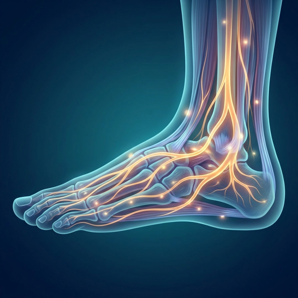
The posterior tibial tendon serves as the primary dynamic stabilizer of the medial longitudinal arch and controls hindfoot inversion during gait. When this tendon fails, the foot’s architecture progressively collapses.
The biological epicenter of PTTD pathology lies in what researchers call the zone of hypovascularity—a 14mm corridor spanning from just proximal to the navicular insertion up to the medial malleolus. This region receives markedly diminished blood supply compared to the rest of the tendon.
Poor blood supply is catastrophic for tendon health. Tendons rely on vascular delivery of oxygen, nutrients, and healing cells. Without adequate perfusion, microtrauma accumulates faster than it can be repaired. This avascular zone is the most common site of tendinopathy and rupture in PTTD patients.
This biological reality exposes the fundamental limitation of standard conservative care. Orthotics and physical therapy reduce mechanical load, but they cannot deliver the biological signals needed to repair degenerated tendon tissue in an avascular zone. The tendon simply lacks the vascular infrastructure to recruit healing factors on its own.
If the problem is biological—insufficient blood supply and an absence of healing cells—the solution must also be biological. This is precisely the rationale behind injecting concentrated growth factors or regenerative cells directly into the zone of hypovascularity. Our foot pain regenerative medicine protocol addresses this biological gap with targeted interventions designed for the avascular environment.
The Four Stages of PCFD: A Framework for Understanding Progression
Understanding the four-stage classification helps patients and clinicians communicate effectively about disease severity and appropriate interventions.
Stage I presents as tendinopathy without deformity. The tendon is inflamed and painful, but the arch remains intact. The “too many toes” sign is absent, and single-limb heel raise is painful but possible.
Stage II marks the onset of flexible flatfoot deformity. The tendon has elongated or partially torn, causing the arch to collapse under weight-bearing. When viewed from behind, excess toes become visible lateral to the heel—the classic “too many toes” sign. Single-limb heel raise becomes impossible or severely limited.
Stage III represents rigid flatfoot deformity. The collapsed arch becomes fixed and cannot be passively corrected. Surrounding joints begin developing arthritis.
Stage IV involves ankle involvement with valgus tilt of the ankle joint and tibiotalar arthritis. This stage represents the most severe and functionally debilitating presentation.
A critical problem in PTTD management is diagnostic delay. Most patients present at Stage II or III, meaning significant structural damage has already occurred before treatment begins. This underscores why early recognition and intervention matter profoundly.
Who Gets PTTD? The Classic Patient Profile and Its Treatment Implications
The classic PTTD patient is an obese woman in her sixth decade of life, often with hypertension, diabetes, or a history of steroid exposure. Research indicates that a BMI of 25 kg/m² or higher is present in 81.5% of PTTD patients, and risk factors including hypertension, obesity, and diabetes appear in up to 60% of cases.
This patient profile carries significant clinical implications. These comorbidities increase surgical risk—poor wound healing, infection, and anesthesia complications become more likely, and surgical outcomes may be compromised in this population.
Here lies a counterintuitive insight: the same comorbidities that make this patient a higher surgical risk actually make her a stronger candidate for cellular therapies. These minimally invasive treatments do not require general anesthesia and can be performed in an outpatient setting. Additionally, metabolic conditions such as diabetes impair natural tendon healing, reinforcing the biological rationale for delivering concentrated growth factors directly to the avascular zone.
Conservative Care: What It Does Well and Where It Falls Short
Conservative care remains the foundation of PTTD treatment, and appropriately so. Research demonstrates that 83% of Stage I and II patients can be successfully treated non-operatively with a structured protocol of orthotics, physical therapy, and immobilization over three to four months.
Standard conservative care includes NSAIDs for inflammation, short-term immobilization with a boot or cast, physical therapy featuring eccentric loading protocols, and custom or semi-custom orthotics. Eccentric and concentric progressive resistive exercises are particularly valuable, with studies showing significant reductions in pain and improvements in function for Stage I and II patients.
Modern advances have improved conservative options. Digital scanning and 3D printing now enable creation of highly precise custom orthotics that provide targeted arch support and unload the posterior tibial tendon—a significant advancement over traditional fabrication methods. A 2026 systematic review and meta-analysis confirmed orthotic interventions effectively reduce pain, improve function, and correct pathological kinematics in PCFD patients.
Notably, corticosteroid injections are now used sparingly in PTTD due to documented risk of tendon rupture, particularly in the already-vulnerable avascular zone.
Yet conservative care has a ceiling. Orthotics and exercise reduce mechanical stress and inflammation, but they cannot regenerate degenerated tendon tissue, restore vascularity, or stimulate collagen remodeling in the avascular zone. This biological gap is precisely what cellular therapies aim to fill.
Extracorporeal Shockwave Therapy: The Underutilized Bridge Between Physical Therapy and Biologics
Extracorporeal shockwave therapy (ESWT) represents an adjunctive treatment for chronic PTTD that receives surprisingly little attention despite growing evidence. ESWT delivers acoustic energy to the tendon, stimulating neovascularization (new blood vessel formation), collagen synthesis, and growth factor release—directly addressing the hypovascularity problem.
A 2025 case study demonstrated that a five-session ESWT protocol over one month effectively managed a significant flare-up of chronic PTTD and enabled a patient to overcome a two-month therapeutic plateau. While ESWT is not a standalone cure, it can extend the effectiveness of physical therapy and may reduce the need for more invasive interventions in patients who have plateaued.
ESWT is most commonly discussed in the context of plantar fasciitis and Achilles tendinopathy; its specific application to PTTD remains an emerging and underreported area. High-quality randomized controlled trial data specific to PTTD is limited, and ESWT is best considered within a comprehensive treatment plan rather than as isolated therapy.
Cellular Therapies for PTTD: The Biological Bridge Conservative Care Cannot Build
Cellular therapies are not a replacement for conservative care—they are biological interventions designed to address what conservative care structurally cannot: the repair of degenerated, avascular tendon tissue.
The overarching rationale is straightforward. By delivering concentrated growth factors, regenerative cells, or extracellular signaling molecules directly into the zone of hypovascularity, cellular therapies aim to stimulate the healing cascade that the tendon’s poor blood supply prevents from occurring naturally.
The regulatory reality must be acknowledged upfront: as of 2026, neither PRP, stem cell therapy, nor exosome products are FDA-approved specifically for musculoskeletal tendon conditions. All are considered investigational with mixed but promising evidence.
Platelet-Rich Plasma (PRP): Concentrating the Body’s Own Healing Signals
PRP therapy involves drawing a small amount of the patient’s own blood, processing it in a centrifuge to concentrate platelets three to five times above normal levels, and injecting the concentrate into the damaged tendon.
Concentrated platelets release growth factors—including PDGF, TGF-β, VEGF, and IGF-1—that signal tissue repair, stimulate collagen synthesis, and promote angiogenesis. Because the avascular tendon cannot recruit these healing signals through normal blood flow, delivering them via injection bypasses the vascular deficit.
PRP shows promise for tendinopathy broadly, with some studies demonstrating pain reduction and functional improvement. Because PRP is derived from the patient’s own blood (autologous), the risk of adverse immune reaction is minimal. Unlike corticosteroid injections, which reduce inflammation but carry tendon rupture risk, PRP aims to stimulate repair rather than simply suppress symptoms.
However, PRP is not a one-size-fits-all solution. Preparation protocols vary significantly between providers, affecting platelet concentration and growth factor content. Our same-day PRP injection protocol outlines how standardized preparation and imaging guidance improve treatment consistency and outcomes.
BMAC and Stem Cell Therapy: Delivering Regenerative Cells to the Avascular Zone
Bone Marrow Aspiration Concentrate (BMAC) involves aspirating bone marrow from the patient’s iliac crest, concentrating it to isolate mesenchymal stem cells (MSCs) and growth factors, and injecting the concentrate into the damaged tendon.
MSCs have the capacity to differentiate into tenocytes (tendon cells), secrete anti-inflammatory cytokines, and stimulate local tissue repair—making them theoretically well-suited for tendon regeneration.
A significant research milestone came from Penn State, where researchers successfully isolated and cultured human posterior tibial tendon-derived adult stem cells for the first time. These cells differentiated into tenocytes at 10 weeks—the first such achievement with human PTT stem cells. This represents a significant step toward autologous tendon-specific cellular therapy, where stem cells harvested from the patient’s own tendon tissue might produce cells already programmed to become healthy tendon.
Patients should be aware that many commercial products marketed as “amniotic stem cell” injections have been independently tested and found to contain no viable living stem cells after processing and storage. Transparency about the contents of any amniotic product being offered is essential.
BMAC is generally considered a more complex and costly intervention than PRP, typically reserved for patients who have failed conservative care and PRP, or who are seeking to avoid surgery. Patients considering this approach should review BMAC injection recovery time expectations as part of their treatment planning.
Exosome Therapy: The Next Frontier in Tendon Regeneration
Exosomes are nano-sized extracellular vesicles secreted by cells that carry proteins, lipids, and genetic material capable of instructing recipient cells to change their behavior. MSC-derived exosomes can deliver regenerative signals to tendon cells without the risks associated with direct cell implantation.
Preclinical studies show MSC-derived exosomes can improve collagen production, promote angiogenesis, and reduce inflammation in tendon tissue. Research on tendon stem cell-derived exosomes (TSC-Exos) demonstrates they can regulate inflammation and promote high-quality tendon healing via PI3K/AKT and MAPK/ERK1/2 signaling pathways.
Exosome therapy for PTTD remains in the preclinical and early clinical research phase. It is not yet widely available as a clinical treatment and is not FDA-approved for this indication. Understanding the exosome pipeline helps patients contextualize the current state of cellular therapy—PRP and BMAC are available options now, while exosomes represent where the field is heading. A deeper look at exosome therapy for tendon and cartilage healing explains the mechanisms driving this emerging research.
The FDA Regulatory Reality: What Patients Must Know Before Pursuing Biologics
As of 2026, the FDA has not approved PRP, stem cell therapy, or exosome products specifically for musculoskeletal tendon conditions, including PTTD. These treatments are not illegal or inherently unsafe—they are investigational, meaning the evidence base is still being established.
Treatments can be administered within FDA regulatory frameworks without being FDA-approved for a specific indication. Patients evaluating providers should ask critical questions: Are injections performed under ultrasound or imaging guidance? What specific product is being used, and what is its regulatory classification? Can the provider share published evidence supporting the specific protocol?
Unicorn Bioscience operates within FDA regulatory frameworks and uses imaging-guided injection protocols, positioning the practice as a transparent, compliance-aware provider. The absence of FDA approval does not mean these therapies lack value—it means patients should seek qualified, transparent providers and maintain realistic expectations about outcomes.
When Surgery Becomes Necessary: Understanding the Surgical Landscape
Surgery remains the appropriate treatment for advanced PTTD/PCFD, and cellular therapies are not a replacement for surgical intervention in Stage III and IV disease.
Current surgical approaches do not repair the degenerated tendon itself but instead use tendon transfers (flexor digitorum longus transfer), calcaneal osteotomies to realign the heel, and arthrodesis (joint fusion) for rigid deformities. Surgical outcomes for Stage II PTTD show significant improvements, with SEFAS scores improving by a mean of 12 points and SF-36 bodily pain improving by 28 points at 24 months post-surgery.
However, surgery is not the default answer for the classic PTTD patient. The obese, diabetic, hypertensive woman in her sixth decade faces elevated surgical risks. For patients who are not ideal surgical candidates, cellular therapies may extend the window of functional conservative management and delay or prevent the need for surgery.
Untreated PTTD that progresses without intervention creates degenerative changes in the surrounding joints and compensatory changes at the knee and hip that worsen overall musculoskeletal outcomes—underscoring the importance of active intervention even when surgery is not yet indicated.
The Hypovascularity-First Framework: A New Way to Think About PTTD Treatment Decisions
Rather than asking “what stage am I?” as the primary treatment question, patients and providers should ask: “Is the biological environment of this tendon capable of healing with conservative measures alone?”
If a patient is in the zone of hypovascularity—which virtually all symptomatic PTTD patients are—conservative care alone cannot deliver the healing signals the tendon needs. Biological augmentation becomes the logical next step before surgery.
The treatment decision hierarchy through this framework proceeds as follows: conservative care (load reduction, eccentric exercise, orthotics) → biological augmentation (PRP, BMAC, ESWT) → surgery (tendon transfer, osteotomy, arthrodesis)—with each step justified by the biology, not just the stage.
Cost and access remain real barriers. PRP injections typically cost $500–$2,000 out of pocket, while BMAC and stem cell procedures can range from $5,000–$10,000 or more. Most insurance plans do not cover these treatments, as they are considered investigational.
This framework is not anti-surgery—it is pro-biology. Understanding why the tendon cannot self-repair helps patients make more informed decisions about when to pursue biologics and when surgery is the appropriate path.
Conclusion: Treating the Biology, Not Just the Stage
Posterior tibial tendon dysfunction treatment has historically been framed as a staging problem. The real driver of treatment failure, however, is a biology problem—the 14mm avascular zone that prevents the tendon from healing itself.
The 2020 rename to Progressive Collapsing Foot Deformity reflects a broader understanding that should inform more comprehensive treatment planning. Conservative care remains the foundation and succeeds in 83% of Stage I and II patients. Cellular therapies represent a biological bridge for those who fail conservative care. Surgery is appropriate and effective for advanced disease in suitable candidates.
PRP, BMAC, and exosome therapies are investigational but scientifically grounded. Patients deserve transparent, evidence-informed guidance from qualified providers.
The classic PTTD patient—often told she is “too heavy for surgery” or “not bad enough yet for intervention”—deserves a treatment conversation that accounts for her biology, her comorbidities, and her goals, not just her stage number.
Ready to Explore Cellular Therapy for PTTD? Start with a Personalized Evaluation
Patients diagnosed with PTTD or PCFD who are exploring options beyond standard conservative care may benefit from a comprehensive evaluation to determine whether cellular therapies are appropriate for their specific situation.
Unicorn Bioscience offers imaging-guided injection protocols using ultrasound and X-ray guidance, multiple treatment modalities including PRP, BMAC, and exosomes, personalized treatment planning based on individual patient factors, and same-day treatment availability for qualified candidates.
Virtual and in-person consultations are available across eight locations in Texas, Florida, and New York. Patients are encouraged to bring staging information, imaging, and comorbidity history to the consultation so the clinical team can assess whether biologics are appropriate.
Unicorn Bioscience operates within FDA regulatory frameworks and provides clear, honest guidance about what cellular therapies can and cannot achieve for PTTD.
Contact information: (737) 347-0446 | unicornbioscience.com | Virtual consultation options available.
Schedule Your Consultation Today!