
Peroneal Tendon Injury Non Surgical Options: The Injury-Subtype Decision Framework That Tells You If Conservative Care Will Work for Your Specific Diagnosis in 2026
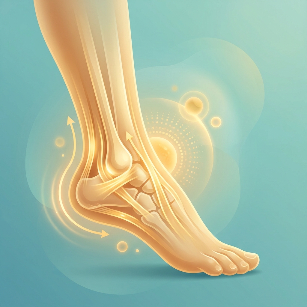
Peroneal Tendon Injury Non-Surgical Options: The Injury-Subtype Decision Framework That Tells You If Conservative Care Will Work for Your Specific Diagnosis in 2026
Introduction: Why Your Peroneal Tendon Diagnosis May Already Be Wrong
Here is a statistic that should give every athlete pause: up to 60% of patients with a peroneal tendon disorder are initially misdiagnosed, most commonly as a simple lateral ankle sprain. That figure is not an outlier. It is supported by a 2025 systematic review and echoed by Cleveland Clinic, which references a study of 40 peroneal tendonitis patients where roughly 60% were mislabeled at first evaluation.
This matters enormously because peroneal tendon injuries are almost always treated as a single condition in both online content and many clinical settings. Patients receive generic protocols, often the same advice given to someone with a routine ankle sprain, when their actual injury may require something entirely different. A protocol that works well for tendinopathy can be useless, or even harmful, for a tendon that is dislocating out of its groove.
This article offers something most content does not: a subtype-stratified decision framework. It maps each category of peroneal tendon injury to the specific non-surgical protocols that fit it, along with realistic, evidence-based outcome expectations. The injury spectrum covered here includes tendinopathy, tenosynovitis, partial tears, complete ruptures, and subluxation/dislocation, each carrying its own conservative care success rate.
This is written for runners, dancers, basketball players, skiers, and active individuals who want to make an informed decision about whether non-surgical care will realistically work for their specific diagnosis before committing to surgery. To be clear, this is not an argument for avoiding surgery when it is genuinely needed. It is a framework for determining when conservative care is appropriate and when it is not.
Understanding the Peroneal Tendon System: Anatomy That Determines Treatment Path
The peroneal system has two primary tendons. The peroneus brevis is shorter and inserts at the base of the fifth metatarsal. The peroneus longus is longer, crossing under the foot to reach the first metatarsal. They sit in different locations, sustain injury through different mechanisms, and respond differently to non-surgical care.
Together, these tendons drive eversion of the foot, support plantarflexion, and provide lateral ankle stabilization. This is why an injury here disrupts balance, push-off power, and the ability to change direction quickly.
A critical structure is the superior peroneal retinaculum (SPR), the band that holds the tendons within the fibular groove behind the ankle. Damage to the SPR is the defining factor in subluxation and dislocation injuries.
The populations most at risk are predictable: endurance runners, dancers, ice skaters, basketball players, skiers, soccer players, and gymnasts, all of whom subject the ankle to repetitive loading or rapid direction changes. Primary risk factors include high foot arches (pes cavus), tight calf muscles, overpronation or excess eversion, inappropriate footwear, rapid increases in training load, and chronic lateral ankle instability.
Anatomy is not academic here. The specific tendon involved, the integrity of the retinaculum, and the degree of displacement directly determine whether non-surgical care can succeed.
The Misdiagnosis Problem: Why Getting the Right Diagnosis Is Step One
The 60% misdiagnosis rate deserves emphasis because it is the single biggest obstacle to effective treatment. Peroneal tendon injuries and lateral ankle sprains share overlapping symptoms: lateral ankle pain, swelling, and tenderness. Without imaging, clinical differentiation is genuinely difficult.
The consequences are real. A patient given ankle sprain protocols (RICE, early mobilization) receives no treatment aimed at the actual tendon pathology. The result can be a chronic injury, recurrent dislocation, or progression to a complete rupture.
Imaging is not interchangeable, and this is where many patients are underserved. According to research published in the Journal of Clinical Medicine, ultrasound achieves 100% sensitivity for peroneal subluxation versus only 66% for MRI, while MRI achieves 100% sensitivity and specificity for peroneal tendon tears. The right imaging modality depends on the suspected injury subtype.
Patients should request imaging that matches their suspected subtype and seek a musculoskeletal specialist or sports medicine physician if their diagnosis was made without any imaging at all. No non-surgical protocol can be properly designed without an accurate diagnosis, making this section the gateway to everything that follows.
The Injury-Subtype Decision Framework: Matching Diagnosis to Non-Surgical Success Rates
Non-surgical success rates vary dramatically by injury subtype. Understanding where a diagnosis falls on the spectrum is the most important factor in choosing a treatment path.
The four primary subtypes covered below are: tendinopathy/tendinitis, tenosynovitis, partial tears, and subluxation/dislocation. Complete ruptures with significant displacement are generally poor candidates for conservative care and are addressed briefly for completeness.
Each subtype section includes the injury definition, the conservative success rate, the recommended non-surgical protocol, a realistic timeline, and the red flags that suggest surgery may be necessary.
Subtype 1: Peroneal Tendinopathy and Tendinitis — The Highest Conservative Success Rate
Tendinopathy refers to degenerative change within the tendon substance without a significant structural tear. Tendinitis refers to inflammatory irritation. Both are the most common peroneal presentations and the best candidates for conservative care.
The success rate is encouraging. The large majority of tendinopathy cases resolve with structured non-surgical management. Mild cases typically resolve within 3 to 4 weeks, and moderate cases within 6 to 8 weeks, per Cleveland Clinic.
The core protocol includes activity modification, RICE during the acute phase, NSAIDs for pain and inflammation, immobilization in a walking boot for 2 to 6 weeks depending on severity, and progressive physical therapy.
Eccentric loading is the evidence-backed cornerstone. Slow eccentric eversion exercises, emphasizing the controlled lowering phase, have the strongest clinical evidence for tendon remodeling and are considered the gold standard for peroneal tendinopathy rehabilitation. Patients dealing with persistent symptoms may also benefit from reviewing chronic tendonitis treatment options that go beyond standard protocols.
Orthotics and footwear also matter: a lateral heel wedge offloads the peroneal insertion, heel-drop shoes of 10 to 12mm reduce strain, and deep heel cup insoles address biomechanical root causes.
Red flags for surgical consideration include failure to respond after 3 to 6 months of structured conservative care, worsening symptoms despite compliance, or imaging showing progressive structural change.
The most common mistake is complete rest followed by a sudden return to activity. Tendons heal through progressive loading, and premature return to sport is the leading cause of chronic, recurring tendinopathy.
Subtype 2: Peroneal Tenosynovitis — Treating the Sheath, Not Just the Tendon
Tenosynovitis is inflammation of the tendon sheath (the synovial lining) surrounding the peroneal tendons. It can occur independently or alongside tendinopathy.
This condition requires a slightly different approach because the inflammatory process in the sheath must be addressed directly, not just the tendon loading pattern. The conservative protocol includes immobilization in a walking boot, NSAIDs, activity modification, and physical therapy targeting both sheath inflammation and the underlying biomechanical contributors.
An important targeted option is ultrasound-guided corticosteroid injection into the peroneal tendon sheath. A PMC/NIH study found this approach safe when performed correctly, with no instances of tendon rupture, infection, or soft-tissue atrophy. The critical distinction is technique: the documented rupture risk associated with corticosteroids is significantly higher with blind (non-guided) injection. Ultrasound guidance is the safety factor that changes the risk profile.
Tenosynovitis generally responds well to conservative management, particularly when treated early, before chronic thickening of the sheath develops.
Red flags include persistent fluid accumulation on repeat ultrasound, failure to respond within 6 to 8 weeks, or the development of a longitudinal split tear within the sheath.
Subtype 3: Partial Peroneal Tendon Tears — Where Conservative Care Becomes Conditional
Partial tears include longitudinal split tears (most common in peroneus brevis) and partial-thickness tears, existing on a spectrum from minor fraying to near-complete disruption.
Conservative success here is conditional. Small partial tears with minimal displacement often respond well. Larger partial tears with significant fiber disruption are far less predictable.
For appropriate candidates, the protocol involves extended immobilization (walking boot for 4 to 6 weeks), followed by structured phased rehabilitation with careful progressive loading:
- Phase 1 (weeks 0 to 2, acute): RICE and immobilization
- Phase 2 (weeks 2 to 4): isometric and concentric strengthening, orthotics
- Phase 3 (weeks 4 to 6): eccentric eversion exercises, single-leg balance
- Phase 4 (week 6 to 8 and beyond): sport-specific agility when eccentric strength reaches at least 90% of the unaffected side
Proprioceptive rehabilitation is critical but often overlooked. Recurrent ankle sprains are linked to poor muscle firing and balance, and each sprain can further damage peroneal tendon fibers.
The tendon involved also matters. Peroneus brevis tears at the fibular groove are more common and more amenable to conservative care, while peroneus longus tears at the cuboid tunnel can be more complex. Patients with adjacent foot and ankle tendon involvement may also want to explore posterior tibial tendon dysfunction treatment as a related condition that can complicate recovery.
Red flags for surgical referral include tears involving more than 50% of the tendon cross-section, significant retraction of torn edges, or failure to progress in a structured 3 to 6 month conservative program. Notably, a Foot & Ankle Clinics systematic review found that no single surgical strategy showed superior functional outcomes, reinforcing the value of a thorough conservative trial first.
Subtype 4: Peroneal Tendon Subluxation and Dislocation — The Most Nuanced Conservative Decision
Subluxation and dislocation involve displacement of the peroneal tendons out of the fibular groove due to damage or laxity of the SPR. Peroneal tendon dislocation accounts for only 0.3 to 0.5% of all ankle traumas, yet it is frequently misdiagnosed as a lateral ankle sprain.
The conservative evidence is meaningful but nuanced. A PMC/NIH systematic review found that a non-weight-bearing cast worn for at least 6 weeks was successful in 5 of 6 patients. A separate systematic review found that a 4 to 6 week below-knee plaster cast prevented recurrent dislocation in 62 to 83% of patients. By contrast, tape fixation alone showed only a 40% success rate, making immobilization quality a decisive variable.
Best candidates for conservative management are those with a first-time acute dislocation, an intact SPR, a low-demand activity level, and no associated tendon tear.
Poor candidates include athletes in high-demand sports (skiing, basketball, soccer), those with recurrent dislocation, significant SPR disruption, or an associated partial or complete tear. Some authors advocate for early surgical repair in competitive athletes even for an initial dislocation.
The conservative protocol is immediate non-weight-bearing immobilization in a below-knee cast or boot, strict activity restriction for the full 6-week period, and graduated physical therapy with proprioceptive retraining.
Red flags include any recurrence of dislocation during or after treatment, persistent instability on examination, or imaging showing complete SPR disruption.
The Standard Non-Surgical Protocol: A Phase-by-Phase Rehabilitation Roadmap
This roadmap applies across subtypes, with the subtype-specific modifications noted above. It fills the gap left by content that mentions only “RICE and physical therapy” without specifics.
- Phase 1, Acute Management (weeks 0 to 2): RICE, NSAIDs, immobilization in a walking boot or cast depending on subtype, and activity modification to eliminate aggravating movements.
- Phase 2, Subacute Strengthening (weeks 2 to 4): Transition to protected weight-bearing, begin isometric eversion and plantarflexion exercises, introduce orthotics or footwear modifications, and begin gastroc-soleus stretching per MGH physical therapy guidelines.
- Phase 3, Progressive Loading (weeks 4 to 6): Introduce eccentric eversion exercises as the primary remodeling stimulus, progress to single-leg balance and proprioceptive drills, continue orthotics, and begin low-impact cross-training (swimming, cycling).
- Phase 4, Return to Sport (week 6 to 8 and beyond): Sport-specific agility and direction-change drills, returning to full training when eccentric eversion strength reaches at least 90% of the unaffected side, per Medscape’s 2026 guidance.
Structured progressive loading with clear milestones rebuilds confidence alongside physical capacity. Chronic or long-standing cases may require 3 to 6 months of structured rehabilitation. The standard of care typically spans 3 to 6 months before surgery is considered, which is key information for anyone trying to understand when surgical referral becomes appropriate. Athletes managing multiple musculoskeletal issues concurrently may benefit from reviewing a sports medicine regenerative treatment protocol to understand how these approaches integrate.
Footwear and Orthotics: The Biomechanical Foundation That Most Protocols Skip
Footwear and orthotics are not optional add-ons. Peroneal tendon overload is almost always driven by biomechanical factors that will continue driving injury unless addressed directly.
- Lateral heel wedge: offloads the peroneal insertion by reducing the mechanical demand on the eversion muscles.
- Heel-drop shoes (10 to 12mm): reduce Achilles and peroneal strain by placing the foot in a more favorable gait position.
- Deep heel cup insoles: improve rearfoot control and reduce excessive subtalar motion that overloads the tendons.
- Custom orthotics for pes cavus: high arches place the foot in a supinated position that dramatically increases peroneal stress, making arch-accommodating orthotics particularly important.
Footwear red flags include worn-down lateral heel counters, zero-drop minimalist shoes, and shoes with insufficient lateral support. Patients should work with a physical therapist or podiatrist for a formal gait analysis before selecting orthotics, since generic off-the-shelf options may not address individual movement patterns.
When Conservative Care Plateaus: Emerging Non-Surgical Options for 2026
When foundational conservative care (immobilization, physical therapy, orthotics) has been properly implemented for 6 to 12 weeks but progress stalls, two emerging modalities become relevant: PRP (Platelet-Rich Plasma) injections and Extracorporeal Shockwave Therapy (ESWT). These are evidence-supported adjuncts that can extend the effectiveness of conservative care, not first-line replacements.
PRP Therapy for Peroneal Tendon Injuries: What the 2024 to 2025 Evidence Shows
PRP is derived from the patient’s own blood, concentrated to deliver a high dose of growth factors (PDGF, TGF-beta, VEGF) directly to the injury site, stimulating tendon cell proliferation and collagen synthesis.
Institutional validation is significant here. Mass General Brigham Sports Medicine explicitly lists peroneal tendon pathology as a candidate condition for PRP, alongside patellar and Achilles conditions. Meta-analyses from 2024 and 2025 indicate PRP can enhance outcomes when used alongside physical therapy, with better healing rates and less pain at follow-up versus standard conservative care alone.
PRP is increasingly preferred over corticosteroids for peroneal pathology because corticosteroids carry a documented rupture risk, while PRP works with the body’s healing mechanisms. The best candidates are patients with tendinopathy or partial tears who have completed 6 to 12 weeks of structured care without adequate response and want a non-surgical option before considering surgery. Understanding the full scope of PRP injection with ultrasound guidance is important for patients evaluating this option, as precision delivery is a key safety and efficacy factor. Ultrasound guidance is essential for precise delivery.
As of 2026, PRP is not FDA-approved specifically for orthopedic tendon conditions, but substantial clinical evidence supports its safety and efficacy when administered by qualified providers within FDA regulatory frameworks. It is not appropriate for complete ruptures with significant displacement or acute dislocations requiring structural repair.
Extracorporeal Shockwave Therapy (ESWT): The 2025 International Consensus and What It Means for Peroneal Tendon Patients
ESWT delivers acoustic pressure waves to tendon tissue, stimulating neovascularization, collagen synthesis, and pain modulation. It is a non-invasive, office-based procedure.
A 2025 international consensus from a panel of 41 rehabilitation and orthopedic experts across 13 countries established recommendations for ESWT use in sports medicine tendinopathy. A major practical advantage is that ESWT typically allows patients to continue training during treatment, a meaningful benefit for competitive athletes.
The best candidates are those with chronic peroneal tendinopathy (symptoms beyond 3 months) that has not responded adequately to standard care. A typical course is 3 to 5 sessions spaced 1 to 2 weeks apart. Some protocols combine ESWT to stimulate the tissue environment with PRP to deliver growth factors, though this combination should be discussed with a qualified provider based on individual presentation.
The Corticosteroid Injection Debate: When It Helps and When It Harms
Corticosteroid injections are commonly offered and can provide meaningful short-term pain relief, so they should not be dismissed outright. However, corticosteroids carry a documented risk of promoting tendon rupture, which is why they are generally considered a last-resort conservative option for peroneal pathology specifically.
The safety distinction lies in technique. As noted, ultrasound-guided injection into the tendon sheath has been shown safe when performed correctly, with no reported ruptures, infections, or soft-tissue atrophy. Rupture risk is significantly higher with blind injection directly into or adjacent to the tendon substance. Patients should ask specifically whether ultrasound guidance will be used.
The appropriate use case is ultrasound-guided injection into the sheath (not the tendon itself) for tenosynovitis that has not responded to initial measures, used as a bridge to enable participation in physical therapy. PRP is increasingly preferred as a next-step injection because it does not carry the rupture risk. Before consenting to any corticosteroid injection, patients should have an informed conversation about the injection technique, guidance method, and target. Patients who want to understand the full range of ultrasound-guided injection for joint pain can review how this approach applies across musculoskeletal conditions.
When Non-Surgical Care Is Not Enough: Recognizing the Surgical Threshold
This section is not about discouraging surgery when it is genuinely indicated. It is about recognizing the evidence-based threshold so patients neither rush into surgery nor delay it when necessary.
The standard of care typically requires 3 to 6 months of structured, compliant conservative care before surgery is considered. Patients who have only tried 2 to 3 weeks of rest have not yet exhausted their options.
Clinical indicators that surgery may be necessary include complete rupture with significant displacement, recurrent dislocation after adequate immobilization, a partial tear involving more than 50% of tendon cross-section, persistent instability on examination, or failure to progress after 3 to 6 months of structured care including advanced options like PRP and ESWT.
There is a high-demand athlete exception: athletes in skiing, basketball, soccer, and similar sports have higher failure rates with conservative management for dislocation, and some authors advocate early surgical repair even for initial dislocations in this population.
Surgical complications, including sural nerve injury, chronic lateral ankle pain, restricted range of motion, and infection, are legitimate reasons to exhaust non-surgical options first. Given that no single surgical strategy has shown superior functional outcomes for peroneal tendon tears, maximizing conservative care is worthwhile. Patients told surgery is immediately necessary without a structured conservative trial should seek a second opinion from a foot and ankle or sports medicine specialist.
Preventing Recurrence: The Long-Term Management Strategy
Recurrence is common and largely preventable. Recovery is not complete at return to sport.
- Ongoing eccentric strengthening: continue a maintenance eccentric eversion program 2 to 3 times per week indefinitely.
- Proprioceptive training: single-leg balance, wobble board, and perturbation drills address the neuromuscular deficits that predispose to re-injury.
- Training load management: follow the 10% rule for weekly progression and build in recovery weeks.
- Footwear maintenance: replace running shoes every 300 to 500 miles and continue using appropriate orthotics.
- Psychological readiness: graduated exposure with clear performance milestones rebuilds confidence and reduces avoidance behavior.
- Annual check-ins: athletes with a significant prior injury benefit from periodic evaluation so minor flares are caught before they become major setbacks.
Conclusion: A Decision Framework for Peroneal Tendon Injury
The central insight is straightforward: peroneal tendon injuries are not one condition. Non-surgical success rates vary dramatically by subtype, from high success in tendinopathy and tenosynovitis, to conditional outcomes in partial tears, to genuinely nuanced decisions in subluxation and dislocation.
Because up to 60% of these injuries are misdiagnosed, the first and most important step is obtaining an accurate diagnosis with appropriate imaging. From there, the decision framework follows a clear hierarchy: accurate diagnosis, then a subtype-matched conservative protocol, then foundational rehabilitation (phased physical therapy, orthotics, footwear), then advanced options like PRP and ESWT when progress plateaus, and finally the surgical threshold only after 3 to 6 months of structured, compliant care.
Individual factors including activity level, sport demands, age, injury severity, and personal goals all shape which options are most appropriate, which is why a personalized assessment is essential. Armed with this framework, patients can ask sharper questions about imaging, injection guidance, and rehabilitation specifics, and make decisions aligned with their recovery goals. Regenerative options like PRP and ESWT are expanding the non-surgical toolkit in 2026, offering meaningful choices for patients who have plateaued on foundational care.
Ready to Explore Personalized Non-Surgical Options for a Peroneal Tendon Injury?
For patients who, based on this framework, recognize themselves as candidates for advanced non-surgical options, Unicorn Bioscience is a logical next step. The practice specializes in cellular and regenerative therapies as alternatives to surgery, including PRP delivered with precision ultrasound-guided injection for accurate therapeutic targeting, the same safety advantage emphasized throughout this article.
Unicorn Bioscience builds personalized treatment plans based on individual injury type, inflammation levels, age, and health goals, and offers a multi-modal approach tailored to the specific subtype and stage of peroneal tendon pathology. Both virtual and in-person consultations are available, which is especially helpful for athletes who lack a regenerative medicine specialist locally. In-person evaluation is available across locations in Texas, Florida, and New York.
The goal is informed decision-making with expert guidance, not a commitment to treatment. Patients who want to know whether PRP or another regenerative option is appropriate for their specific peroneal tendon diagnosis can schedule a consultation as the next step in their decision framework. Call (737) 347-0446 or visit unicornbioscience.com to get started.
Schedule Your Consultation Today!
