
Pain on Outer Side of Knee: The 6-Cause Lateral Anatomy Map That Matches Your Exact Symptom Pattern to the Right Treatment
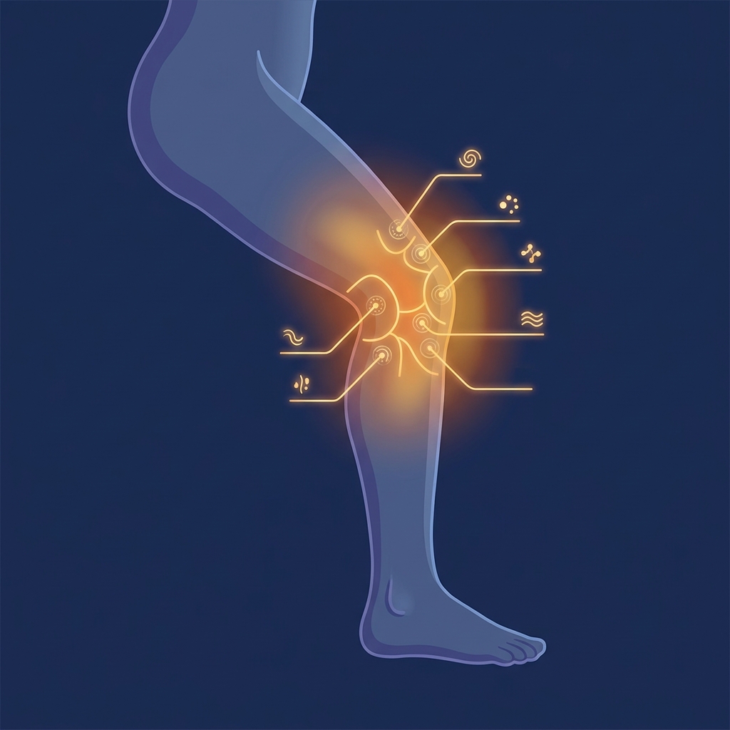
Pain on Outer Side of Knee: The 6-Cause Lateral Anatomy Map That Matches Your Exact Symptom Pattern to the Right Treatment
A competitive runner stops mid-stride, gripping the outside of the knee. A nurse finishes a grueling 12-hour shift and feels a sharp twinge while walking to the parking lot. A remote worker stands up after hours at a desk and winces at a burning sensation along the outer edge of the knee. These scenarios share a common thread: lateral knee pain that demands answers.
Knee pain affects approximately 25% of adults, with prevalence increasing nearly 65% over the past 20 years. This translates to nearly 4 million primary care visits annually in the United States alone. Yet most available resources list the same causes without helping readers differentiate between them based on how their pain actually behaves.
This guide offers something different: a structured symptom-fingerprint system covering six major lateral knee structures, paired with tiered treatment pathways that reflect 2025 and 2026 clinical evidence. Whether the reader is an athlete, a desk worker, a nurse, or a retail employee, this framework applies. For clarity, “lateral” is the medical term for the outer side of the knee, the area furthest from the body’s midline.
Understanding Lateral Knee Anatomy: Why Location Matters for Diagnosis
The outer knee is not a single tissue but a complex region housing multiple overlapping structures. This anatomical complexity explains why pain in this area can have vastly different causes and treatments.
Six key structures warrant attention: the iliotibial (IT) band, the lateral collateral ligament (LCL), the lateral meniscus, the lateral compartment cartilage (relevant to osteoarthritis), the popliteal and biceps femoris tendon complex, and the posterolateral corner (PLC). Each structure produces a distinct pain character, onset pattern, activity trigger, and treatment pathway. Treating the wrong structure wastes time and can worsen outcomes.
The common peroneal nerve also runs along the outer knee and can produce numbness, tingling, and weakness when compressed or injured. This differentiating detail is frequently overlooked in consumer-facing resources.
Additionally, some outer knee pain originates from the hip or lower back through referred pain patterns, reinforcing the importance of accurate diagnosis before beginning treatment.
The 6-Cause Lateral Anatomy Map: Matching Your Symptom Pattern to the Right Diagnosis
Every cause of lateral knee pain produces a unique symptom fingerprint: a combination of pain character, onset pattern, activity triggers, precise location, and associated sensations. Readers should note which description most closely matches their experience, then follow the corresponding treatment pathway.
A summary comparison helps clarify distinctions:
| Cause | Pain Character | Onset Pattern | Key Activity Trigger | Precise Location | Who It Affects Most |
|---|---|---|---|---|---|
| IT Band Syndrome | Burning, dull ache | Gradual with activity | Running, cycling, stairs | Above joint line | Runners, cyclists, desk workers |
| LCL Injury | Sharp, localized | Acute after trauma | Contact sports, falls | Over the LCL | Contact sport athletes |
| Lateral Meniscus Tear | Sharp or aching with clicking | Acute or gradual | Twisting, squatting | At joint line | Pivoting sport athletes, older adults |
| Lateral Compartment OA | Deep, diffuse ache | Gradual over months | Prolonged standing | Broadly across outer knee | Adults over 40, prior injuries |
| Popliteus/Biceps Femoris | Deep ache, posterior | Gradual overuse | Downhill running, sprinting | Back-outer corner | Hill runners, cyclists |
| PLC Injury | Diffuse with instability | Acute after trauma | Rotational activities | Posterolateral corner | Trauma patients, contact athletes |
Self-diagnosis has limits. A healthcare provider should confirm any suspected diagnosis, particularly for moderate-to-severe symptoms.
Cause 1: IT Band Syndrome (ITBS): The Burning Band
IT band syndrome is the most common cause of lateral knee pain, responsible for approximately 10% of all running-related injuries and affecting 5 to 14% of runners. First documented in US Marine Corps recruits in 1975, ITBS has since been diagnosed frequently in long-distance runners, cyclists, skiers, and participants in basketball, soccer, and hockey.
Symptom fingerprint: A dull, burning ache on the outer knee that typically begins after a predictable distance or time into activity. This “pain threshold” phenomenon is characteristic. Pain worsens going downstairs or on downhill terrain and is often absent at rest. Some individuals describe a snapping or rubbing sensation.
Precise location: Pain concentrates at the lateral femoral epicondyle, approximately 2 to 3 cm above the joint line, not at the joint itself.
Who it affects: Runners, cyclists, and team sport athletes are commonly affected. However, ITBS also impacts walkers, desk workers, nurses, and retail workers with weak glutes, poor posture, or worn-out footwear. A 2024 large-cohort study of 76,654 race entrants found higher prevalence in females, younger athletes, those with fewer years of running experience, and those with a history of chronic diseases or allergies.
Current evidence suggests ITBS results from compression of a fat pad beneath the IT band rather than friction alone. This distinction matters for treatment. The widely repeated instruction to “stretch your IT band” is clinically inaccurate. The IT band is dense connective tissue that cannot be meaningfully lengthened with stretching.
Cause 2: Lateral Collateral Ligament (LCL) Injury: The Instability Signal
Symptom fingerprint: Sharp, localized pain on the outer knee, often associated with a sense of instability or giving way. Pain is typically immediate in onset following a specific incident.
Precise location: Pain is felt directly over the LCL, which runs from the lateral femoral epicondyle to the fibular head.
Onset pattern: Acute onset following a direct blow to the inner (medial) side of the knee or a sudden varus stress (knee forced inward). Less commonly, gradual onset occurs from chronic varus loading.
Associated sensations: Swelling, bruising, and stiffness may occur. In severe cases, numbness or tingling in the foot indicates common peroneal nerve involvement.
LCL injuries are graded: Grade I (mild sprain), Grade II (partial tear), and Grade III (complete tear), each with different recovery expectations. Isolated LCL injuries are less common than combined posterolateral corner injuries. If instability is prominent, PLC involvement should be evaluated.
Cause 3: Lateral Meniscus Tear: The Locking, Clicking Pain
Symptom fingerprint: Sharp or aching pain at the joint line (the crease where the femur meets the tibia), often accompanied by clicking, locking, or catching sensations. Pain worsens with twisting, squatting, or pivoting.
Precise location: Pain is felt at the lateral joint line, lower and more precisely at the joint crease than ITBS pain.
Onset pattern: Acute onset following a twisting injury with the foot planted, or gradual onset in older adults through degenerative tearing.
Only the outer portion of the lateral meniscus has a blood supply, meaning most tears in the inner zone do not heal on their own and may require intervention.
Distinguishing from ITBS: Meniscal pain is at the joint line and provoked by rotation; ITBS pain is above the joint line and provoked by repetitive linear motion. For patients exploring non-surgical options, meniscus repair with stem cell therapy has emerged as a promising alternative to traditional surgical intervention.
Cause 4: Lateral Compartment Osteoarthritis: The Deep, Weather-Sensitive Ache
Symptom fingerprint: Deep, diffuse aching pain on the outer knee; stiffness after rest (especially morning stiffness lasting less than 30 minutes); pain that worsens with prolonged weight-bearing and improves with rest. Some individuals report worsening in cold or damp weather.
Precise location: Pain is felt broadly across the outer knee compartment rather than at a specific point.
Onset pattern: Gradual, progressive onset over months to years with no single precipitating event.
Globally, knee osteoarthritis affects approximately 654 million people aged 40 and older. Each extra pound of body weight adds approximately 4 pounds of pressure to the knee, and obese individuals face a fourfold increased risk. Lateral compartment OA is less common than medial compartment OA but is associated with varus (bow-legged) alignment.
Cause 5: Popliteus Tendon and Biceps Femoris Tendinopathy: The Overlooked Overuse Injuries
These two tendons are frequently overlooked causes of outer knee pain, often misdiagnosed as ITBS.
Popliteus tendon symptom fingerprint: Pain at the back-outer corner of the knee that worsens with downhill running or walking. It may feel like a deep ache rather than a surface burn and is often provoked by resisted internal rotation of the tibia.
Biceps femoris tendon symptom fingerprint: Pain at the outer back of the knee where the hamstring attaches to the fibular head. It worsens with hamstring loading (sprinting, hill climbing) and may be associated with a snapping sensation.
Distinguishing from ITBS: Location is more posterior; pain is provoked by different movements; the characteristic pain threshold pattern of ITBS is absent.
Cause 6: Posterolateral Corner (PLC) Injury: The Often-Missed Diagnosis
The PLC is a complex of structures (LCL, popliteus tendon, popliteofibular ligament, and others) that together stabilize the outer knee against varus and rotational forces. PLC injuries are frequently confused with isolated LCL sprains and are underdiagnosed in emergency and primary care settings.
Symptom fingerprint: Outer knee pain combined with significant instability, particularly a varus thrust (the knee buckling outward during walking). Pain occurs with activities requiring rotational stability. Associated peroneal nerve symptoms (foot drop, numbness along the outer shin) may be present.
Red flag: If outer knee pain is accompanied by significant instability, inability to bear weight, or neurological symptoms, urgent evaluation is required.
Red Flags: When Outer Knee Pain Requires Urgent Medical Attention
Certain symptoms require same-day or emergency evaluation: inability to bear weight, gross deformity, numbness or pallor in the foot, severe instability, or signs of joint infection (fever, significant warmth, redness, extreme swelling). These may indicate fracture, vascular injury, complete ligament rupture, septic arthritis, or significant nerve damage.
Foot drop (inability to lift the foot) following outer knee trauma is a neurological emergency indicating common peroneal nerve injury.
Even without red flags, persistent pain lasting more than 2 to 3 weeks, pain that worsens despite rest, or pain that significantly limits daily function warrants medical evaluation.
Getting the Right Diagnosis: What to Expect at Your Appointment
A typical diagnostic encounter for lateral knee pain includes history-taking (onset, activity triggers, pain character, prior injuries), physical examination (palpation, range of motion, special tests), and imaging selection.
Key physical examination tests include the Noble compression test and Ober test for ITBS; varus stress test for LCL; McMurray and Thessaly tests for meniscal tears; and the dial test for PLC.
X-rays are first-line to rule out fractures and assess joint space narrowing. MRI is the gold standard for soft tissue pathology. Ultrasound is useful for dynamic assessment of the IT band, bursae, and tendons. Ultrasound-guided injections, such as those used at Unicorn Bioscience, improve accuracy of therapeutic delivery compared to landmark-guided injections.
Readers should bring a symptom log to their appointment: when pain started, what makes it better or worse, its character, and its precise location.
Tiered Treatment Pathways: From Conservative Care to Regenerative Options
Most lateral knee pain responds to conservative care. Regenerative and procedural options are appropriate when conservative measures fail after an adequate trial, typically 6 to 12 weeks.
Tier 1: Conservative Care: The Foundation for All Six Causes
Rest and activity modification: Relative rest (reducing aggravating activities) is preferred over complete rest, which may worsen outcomes.
Ice and compression: Appropriate for acute pain and swelling; 15 to 20 minutes several times daily in the acute phase.
NSAIDs: Appropriate for short-term pain and inflammation management, though long-term use carries gastrointestinal and cardiovascular risks.
Physical therapy: The cornerstone of conservative treatment for all six causes. Protocols differ by diagnosis. ITBS requires hip abductor strengthening, LCL injuries require stability work, and meniscal tears require quadriceps and hamstring balance. A 2024 systematic review found that hip abductor strengthening exercises, possibly augmented by shockwave therapy or manual therapy, are the most effective conservative treatments for ITBS.
Footwear assessment: Worn-out or biomechanically inappropriate footwear is a modifiable risk factor.
Recovery timelines: Mild ITBS typically resolves in 4 to 8 weeks with appropriate treatment. Grade I LCL sprains heal in 2 to 4 weeks. Lateral meniscal tears vary widely. OA is managed long-term rather than cured.
Tier 2: Intermediate Interventions
Corticosteroid injections offer short-term pain relief for ITBS, OA, and tendinopathy, though repeated injections carry risks of tissue weakening. Hyaluronic acid (viscosupplementation) is an evidence-supported option for lateral compartment OA. Bracing and orthotics can shift load away from the affected compartment. Shockwave therapy and dry needling may provide adjunctive benefits.
For desk workers, nurses, and retail workers, ergonomic modifications are important adjuncts to clinical treatment.
Tier 3: Regenerative Medicine: The 2025 and 2026 Evidence Update
Regenerative options are appropriate for patients who have not responded adequately to conservative care or who wish to avoid surgery.
PRP (Platelet-Rich Plasma): A 2025 meta-analysis showed clinically significant improvements in knee pain and function over placebo and corticosteroids at 6 to 12 months, with efficacy influenced by platelet concentration. PRP is derived from the patient’s own blood, minimizing rejection risk.
Stem cell therapy and BMAC (Bone Marrow Aspiration Concentrate): Emerging evidence supports use in OA and ligament injuries. Unicorn Bioscience reports that more than 90% of stem cell patients have not gone on to knee replacement surgery. Learn more about stem cell therapy for knee pain and the latest clinical evidence supporting these approaches.
Exosome therapy: An emerging modality using extracellular vesicles to support cellular communication and tissue regeneration. Patients interested in this option can explore exosome injection for joint pain as part of a comprehensive regenerative protocol.
Genicular artery embolization (GAE): A minimally invasive vascular procedure showing a 99.7% technical success rate with pain reduction of 34 to 39 points on the Visual Analog Scale.
All injections at Unicorn Bioscience are administered under ultrasound or X-ray guidance, improving precision and outcomes. Same-day treatment is available for qualified candidates. Regenerative options are most effective when combined with a structured physical therapy program.
As of 2026, the FDA has not approved stem cell, PRP, or exosome products specifically for orthopedic conditions, but substantial clinical evidence supports safety and efficacy when administered by qualified providers within FDA regulatory frameworks.
Tier 4: Surgical Options
Surgery is rarely required for ITBS and is reserved for cases that fail 6 to 12 months of comprehensive conservative treatment. Lateral meniscus tears may require partial meniscectomy or meniscal repair. Grade III LCL tears and complex PLC injuries often require surgical reconstruction. For severe lateral compartment OA, joint replacement is the surgical endpoint, though studies suggest up to 80% of patients told they need total knee replacement may not actually require surgery. Patients facing this decision may benefit from reviewing whether knee replacement surgery is truly necessary before committing to an invasive procedure.
Prevention: Reducing Risk of Lateral Knee Pain
Hip and glute strengthening: The single most evidence-supported preventive strategy for ITBS. Examples include clamshells, lateral band walks, and single-leg squats.
Training load management: Avoid rapid increases in mileage or intensity.
Footwear: Replace running shoes every 300 to 500 miles.
Address sedentary lifestyle risk: Prolonged sitting weakens the glutes and tightens the hip flexors, increasing lateral knee loading when activity resumes.
Weight management: Maintaining a healthy weight is one of the most impactful preventive measures for OA.
Frequently Asked Questions About Pain on the Outer Side of the Knee
Why does outer knee pain worsen when walking downstairs or downhill? Downhill and stair descent increase IT band compression at the lateral femoral epicondyle. This is a hallmark symptom of ITBS.
Can running continue with outer knee pain? Mild ITBS may allow continued running with reduced volume. Sharp pain, instability, or swelling are signals to stop and seek evaluation.
How long does IT band syndrome take to heal? Mild cases resolve in 4 to 8 weeks; chronic or severe cases may take 3 to 6 months.
What is the difference between IT band pain and meniscus pain? IT band pain is above the joint line, burning in character, and provoked by repetitive linear motion. Meniscal pain is at the joint line, sharp or aching, and provoked by twisting or rotation.
Conclusion: Matching Pain Pattern to the Right Path Forward
Pain on the outer side of the knee is not a single condition but a symptom with six distinct structural causes, each with a unique symptom fingerprint and treatment pathway. Readers who have identified their likely cause should now have a clearer understanding of what conservative steps to take first and when to escalate to professional care.
The 2025 and 2026 evidence supports PRP, regenerative cellular therapies, and minimally invasive procedures as effective alternatives to surgery for appropriate candidates. One outdated myth also deserves correction: stretching the IT band is not the answer. Strengthening the hip and addressing biomechanical contributors is the evidence-based path to lasting relief.
Whether the reader is a competitive runner, a nurse on a 12-hour shift, or a remote worker rediscovering movement, the right diagnosis and the right treatment pathway can restore pain-free function.
Take the Next Step: Explore Regenerative Treatment Options for Lateral Knee Pain
For those who have not found relief through conservative care or who are seeking alternatives to surgery, regenerative medicine offers promising options.
Unicorn Bioscience provides cutting-edge regenerative medicine for orthopedic conditions, with 8 locations across Texas (Austin, Dallas, El Paso, Fort Worth, Houston, San Antonio), Florida (Boca Raton), and New York (Manhattan). Key differentiators include precision imaging-guided injections using ultrasound and X-ray, multiple treatment modalities (PRP, stem cell therapy, BMAC, exosome therapy, hyaluronic acid, peptide therapy), same-day treatment availability for qualified candidates, and personalized treatment planning based on individual patient factors.
To discuss whether regenerative treatment is appropriate for a specific lateral knee condition, schedule a virtual or in-person consultation by calling (737) 347-0446 or visiting unicornbioscience.com.
Schedule Your Consultation Today!
