Pain Knee Squat: The 4-Location Diagnostic Protocol That Tells You Exactly What’s Wrong
Pain Knee Squat: The 4-Location Diagnostic Protocol That Tells You Exactly What’s Wrong
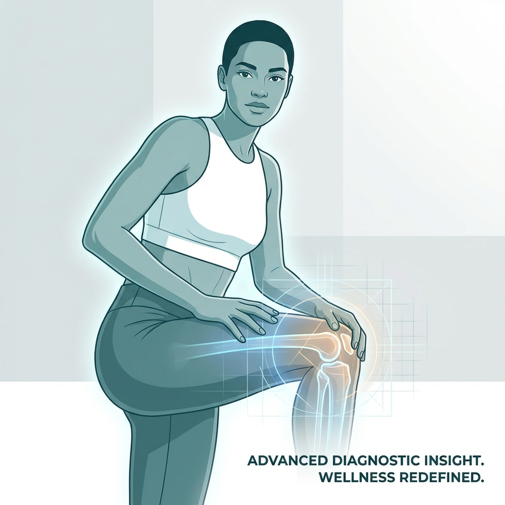
Introduction: Why Knee Pain During Squats Demands a Location-First Diagnosis
Knee pain represents one of the most common musculoskeletal complaints worldwide, with global prevalence ranging from 10% to 60% across populations. In the United States alone, approximately 25% of adults over age 45 report frequent knee pain, and roughly 5% of all primary care visits involve knee-related concerns. For individuals who experience pain specifically during squatting, the frustration compounds quickly because squatting is not merely an exercise. It is a fundamental human movement embedded in daily activities, athletic training, and rehabilitation protocols.
The core problem with most guidance on squat-related knee pain is that it treats the complaint as a single condition. In reality, knee pain during squats represents a family of distinct diagnoses separated by location and movement phase. Generic advice fails because anterior knee pain has different causes than medial pain, and pain at the bottom of a squat indicates different pathology than pain during the descent.
This article introduces a dual-axis diagnostic framework that combines pain location (anterior, medial, lateral, posterior) with squat phase (descent, bottom position, ascent) to create a precise diagnostic grid. Additionally, two underrepresented contributors are addressed: ankle mobility restriction and the distinction between unilateral versus bilateral pain patterns. By the end of this protocol, readers will be equipped to narrow down their likely diagnosis, understand the biomechanical reasoning behind it, and map a clear path from self-care to clinical intervention.
How to Use This Diagnostic Protocol: The Dual-Axis Framework Explained
The diagnostic framework operates on two axes. Axis 1 identifies the anatomical quadrant of pain: anterior (front), medial (inside), lateral (outside), or posterior (back). Axis 2 identifies the squat phase when pain peaks: descent, bottom position at full flexion, or ascent.
To self-assess pain location accurately, performing a controlled bodyweight squat while focusing on the primary area of discomfort is essential. Anatomical landmarks help pinpoint the quadrant: the kneecap indicates anterior pain, the inner joint line indicates medial pain, the outer aspect indicates lateral pain, and the back of the knee indicates posterior pain.
Understanding the three squat phases clarifies what each phase mechanically stresses. The descent phase involves eccentric quadriceps loading and increasing patellofemoral compression. The bottom position, typically at 60 to 90 degrees of knee flexion, creates maximum joint compression and the highest meniscal load. The ascent phase involves concentric quadriceps firing and peak tendon tension.
Notably, patellofemoral joint forces can reach up to 427% of body weight during deep squats, with peak compressive forces occurring between 60 and 90 degrees of knee flexion. This biomechanical reality explains why the bottom position is often the most painful phase for many individuals.
A third diagnostic variable warrants attention: whether pain is unilateral (one knee) or bilateral (both knees). Unilateral pain suggests structural asymmetry, prior injury, or localized pathology. Bilateral pain more often points to systemic overuse, technique errors, or ankle mobility deficits affecting both sides equally.
This framework serves to narrow the differential diagnosis before clinical evaluation, not to replace professional assessment.
Anterior Knee Pain During Squats: The Most Common Quadrant
Anterior knee pain is the most frequently reported location during squatting and accounts for up to 40% of all clinical knee visits. The primary diagnoses within this quadrant include Patellofemoral Pain Syndrome (PFPS), patellar tendinitis, chondromalacia patellae, and fat pad impingement.
Patellofemoral Pain Syndrome (PFPS): The Leading Diagnosis
PFPS involves pain arising from the interface between the patella and the femoral groove, caused by abnormal patellar tracking, excessive compressive forces, or cartilage stress. The condition carries an annual prevalence of approximately 22.7% in the general adult population, with women affected at nearly twice the rate of men. Among athletes, PFPS accounts for 33% of knee injuries in females and 18% in males.
The classic presentation includes diffuse retropatellar or peripatellar pain that worsens during squatting, stair climbing, and prolonged sitting with knees bent. This latter symptom is often called the “movie sign.”
The diagnostic power of the squat test is substantial. According to clinical practice guidelines, the presence of anterior knee pain during a squat is approximately 91% sensitive and 50% specific for PFPS, making it the single best clinical diagnostic test for this condition.
Phase correlation reveals that PFPS pain typically peaks at the bottom of the squat and during the descent phase when patellofemoral compression is highest. Biomechanical contributors include weak gluteus medius and poor gluteus maximus activation. Research demonstrates that individuals with PFPS show significantly reduced gluteus medius activity during squats compared to healthy controls. Additionally, anterior tibial translation past the toes serves as the primary aggravating technique factor.
Approximately 25% of recreational athletes with PFPS stop participating in sports due to pain, underscoring the importance of early intervention. For patients with persistent symptoms, patellofemoral arthritis treatment options may provide relief when conservative measures fall short.
Patellar Tendinitis and Chondromalacia: Related but Distinct
Patellar tendinitis, commonly called “jumper’s knee,” produces pain localized to the inferior pole of the patella or the patellar tendon itself. This pain typically worsens during the ascent phase when the patellar tendon is under maximum tensile load.
Chondromalacia patellae involves softening and degeneration of the cartilage on the underside of the patella. Pain is often described as a grinding or crunching sensation during knee flexion. Phase correlation mirrors PFPS, with symptoms worst at the bottom of the squat.
Fat pad impingement, known as Hoffa’s syndrome, produces pain just below and around the kneecap. This condition is often worsened at full knee extension during the ascent and represents a frequently overlooked anterior pain source.
A key differentiator: patellar tendinitis pain is point-tender at the tendon insertion, while PFPS pain is diffuse and retropatellar. Both conditions can coexist.
Importantly, avoiding squats due to anterior pain often leads to quadriceps atrophy and worsened patellar tracking. This fear-avoidance pattern is counterintuitive but clinically significant. Patients with chronic tendonitis affecting the patellar tendon may benefit from targeted regenerative approaches rather than prolonged rest.
Medial Knee Pain During Squats: Inside-of-the-Knee Diagnoses
Medial knee pain during squatting has a distinct differential from anterior pain and requires different assessment and treatment approaches. Primary diagnoses include medial meniscus tear, MCL sprain, pes anserine bursitis, medial plica syndrome, and saphenous nerve entrapment.
Medial Meniscus Tears and MCL Sprains
Medial meniscus tears produce pain along the medial joint line, often accompanied by joint-line tenderness on palpation. Deep squatting to the bottom position typically provokes sharp or catching pain as the meniscus is compressed between the femur and tibia.
Phase correlation indicates that medial meniscus pain is most pronounced at the bottom of the squat at maximum flexion. Accompanying symptoms may include a click, pop, or sensation of locking. These are red-flag symptoms warranting immediate clinical evaluation.
MCL sprains produce medial pain that is more diffuse and often associated with a history of valgus stress or twisting mechanism. Pain occurs during descent when the knee is under valgus load.
Both conditions present almost exclusively as unilateral pain. Bilateral medial pain should prompt consideration of systemic causes such as osteoarthritis or inflammatory arthritis.
Pes Anserine Bursitis and Medial Plica Syndrome
Pes anserine bursitis is a frequently overlooked cause of medial knee pain, characterized by tenderness 5 to 7 centimeters below the anteromedial joint line. This location distinguishes it from meniscal pain, which occurs at the joint line itself. The condition is common in runners, individuals with obesity, and those with knee osteoarthritis.
Pain from pes anserine bursitis typically worsens during both descent and ascent phases when the pes anserine tendons are under load. Symptoms may mimic medial meniscus tear on clinical presentation, requiring imaging for differentiation.
Medial plica syndrome involves irritation of the medial synovial fold, producing a snapping or clicking sensation along the medial aspect of the knee during flexion and extension cycles. Symptoms are most pronounced during the descent phase.
Saphenous nerve entrapment represents a rare but clinically important differential. This condition produces medial knee pain with a burning or tingling quality rather than mechanical pain and does not follow joint-line or tendon patterns.
Lateral Knee Pain During Squats: Outside-of-the-Knee Diagnoses
Lateral knee pain during squatting is most commonly associated with IT band syndrome in active individuals and lateral compartment osteoarthritis in older adults. Primary diagnoses include iliotibial band syndrome (ITBS), lateral meniscus tear, lateral compartment osteoarthritis, and biceps femoris tendinopathy.
Iliotibial Band Syndrome: The Most Common Lateral Diagnosis
ITBS involves inflammation where the iliotibial band crosses the lateral femoral epicondyle. It is the most common cause of lateral knee pain in athletes, accounting for approximately 12% of all running injuries, and is commonly triggered by repetitive squatting movements.
The biomechanical mechanism centers on the ITB making contact with the lateral epicondyle at approximately 30 degrees of knee flexion. This means ITBS pain is characteristically provoked during the descent phase as the knee passes through this impingement zone.
The classic presentation includes sharp, burning lateral knee pain worsened by repetitive knee flexion and extension. Pain peaks during the descent phase at approximately 30 degrees of flexion and may diminish at the bottom of a deep squat beyond the impingement zone. This pattern serves as a useful diagnostic clue.
Contributing factors include hip abductor weakness, excessive hip adduction during squatting (knee caving inward), and training volume spikes. ITBS is typically unilateral; bilateral presentation suggests a systemic training error or biomechanical issue.
Lateral Meniscus Tears and Lateral Compartment OA
Lateral meniscus tears produce lateral joint-line pain most severe at the bottom of the squat. Accompanying symptoms may include clicking, locking, or giving-way sensations, which are red-flag symptoms requiring clinical evaluation. Patients exploring knee meniscus cellular therapy candidacy may find regenerative options appropriate depending on tear severity and location.
Lateral compartment osteoarthritis produces diffuse lateral pain that worsens progressively throughout the squat movement. It is more common in older adults and those with varus knee alignment. Pain is typically bilateral but asymmetric.
Biceps femoris tendinopathy produces pain at the lateral aspect of the knee near the fibular head, worsened during the ascent phase when the biceps femoris is under concentric load.
Posterior Knee Pain During Squats: The Most Overlooked Quadrant
Posterior knee pain during squatting is the least common presentation but is consistently underrepresented in educational content. When it does occur, the differential is specific and clinically significant. Primary diagnoses include posterior meniscal horn tears, Baker’s cyst, hamstring tendinopathy, gastrocnemius tendinopathy, popliteus tendinopathy, and PCL injury.
Baker’s Cyst and Posterior Meniscal Horn Tears
A Baker’s cyst is a fluid-filled sac in the popliteal fossa that causes a sensation of tightness, fullness, or aching at the bottom of the squat when the cyst is compressed. It is often secondary to an underlying intra-articular pathology such as a meniscal tear or osteoarthritis.
Baker’s cyst discomfort is most pronounced at the bottom of the squat at full flexion and may cause a blocked sensation limiting squat depth.
Posterior meniscal horn tears involve the most commonly torn regions of the menisci. Pain is localized to the posterior joint line and is worst at the bottom of the squat under maximum compressive load. These tears are often missed on physical exam and require MRI for confirmation.
A critical clinical pearl: a Baker’s cyst that ruptures can cause sudden posterior knee and calf pain mimicking a deep vein thrombosis. This presentation warrants urgent evaluation.
Hamstring, Gastrocnemius, and Popliteus Tendinopathy
Distal hamstring tendinopathy produces posterior knee pain during the descent phase as the hamstrings eccentrically load. Pain is often described as a deep ache behind the knee.
Gastrocnemius tendinopathy produces pain at the posterior knee near the medial or lateral gastrocnemius origin, worsened during the ascent phase when the gastrocnemius contributes to knee flexion control.
Popliteus tendinopathy is a frequently overlooked cause of posterolateral knee pain. The popliteus muscle stabilizes the knee during the descent phase, and pain is provoked during downhill squatting or loaded descent movements.
PCL injuries produce posterior knee pain and instability. Squatting with significant load can provoke posterior tibial sag and pain. This is a red-flag diagnosis requiring imaging. Cellular therapy for ligament tears represents a non-surgical option worth evaluating for appropriate candidates.
The Hidden Contributors: Ankle Mobility and Unilateral Pain Patterns
Two clinically significant contributors to squat knee pain are consistently underrepresented in educational content.
Ankle Dorsiflexion Restriction: The Upstream Cause of Knee Pain
Common causes of ankle dorsiflexion restriction include tight gastrocnemius and soleus complex, prior ankle sprains with scar tissue, and bony ankle impingement. Patients with a history of ankle sprains may benefit from ankle sprain regenerative treatment to address residual scar tissue contributing to restricted dorsiflexion.
The knee-to-wall test provides a simple self-assessment method. Less than 4 inches of clearance suggests clinically significant restriction.
Elevating the heels during squatting using a heel wedge or Olympic lifting shoes reduces anterior tibial translation and can immediately reduce anterior knee pain. This modification serves both diagnostic and therapeutic purposes.
Ankle restriction is often bilateral and symmetric, making it a common cause of bilateral anterior knee pain during squats.
Unilateral vs. Bilateral Pain: What the Pattern Reveals
Unilateral knee pain during squatting strongly suggests a structural or localized cause: meniscal tear, ligament injury, focal osteoarthritis, bursitis, or prior trauma. Leg length discrepancy and asymmetric hip or ankle mobility also warrant consideration.
Bilateral knee pain during squatting more often points to systemic overuse, technique errors, ankle mobility restriction, or inflammatory and degenerative conditions affecting both joints.
New-onset unilateral pain in a previously pain-free squatter should raise suspicion for an acute structural injury and warrants prompt evaluation. Gradual bilateral onset more commonly reflects overuse or technique-related issues.
Red Flag Symptoms: When Knee Pain During Squats Requires Immediate Evaluation
Certain symptoms require immediate clinical attention:
- Locking or catching: The knee physically locks and cannot be fully extended, suggesting a displaced meniscal tear or loose body.
- Giving way or instability: The knee buckles during squatting or walking, suggesting ligamentous injury or significant cartilage loss.
- Significant acute swelling: Rapid joint effusion within hours of injury suggests hemarthrosis from ACL tear, fracture, or severe meniscal tear.
- Night pain: Pain that wakes the patient from sleep may indicate inflammatory arthritis, infection, or neoplasm.
- Inability to bear weight: Suggests fracture or complete ligament rupture.
- Posterior knee pain with calf swelling: May indicate a ruptured Baker’s cyst or DVT.
- Fever with knee pain: Suggests septic arthritis, a medical emergency.
If any red flag is present, seeking clinical evaluation rather than attempting self-management is essential.
The Treatment Continuum: From Self-Care to Regenerative Cellular Therapy
The appropriate treatment depends on the specific diagnosis identified through the location-phase framework, not a one-size-fits-all approach.
Level 1: Self-Care and Conservative Management (Weeks 1 to 4)
For mild overuse-related pain without red flag symptoms, conservative measures include relative rest and activity modification, ice application for acute flares, and short-term anti-inflammatory use under physician guidance.
Ankle mobility work addresses dorsiflexion restriction, while hip strengthening targets gluteus medius and gluteus maximus activation. Technique corrections include heel elevation, reducing squat depth to stay above 60 degrees of flexion, and cueing knee tracking over the second toe.
Mild cases may resolve in 2 to 4 weeks with consistent self-care. Failure to improve within 4 to 6 weeks warrants clinical evaluation.
Level 2: Physical Therapy and Clinical Rehabilitation (Weeks 4 to 12)
For persistent pain beyond 4 to 6 weeks, physical therapy provides individualized assessment of movement patterns and progressive loading protocols. Patellar taping and knee sleeves can provide short-term pain relief for PFPS. Corticosteroid injections are appropriate for acute inflammatory conditions but do not address underlying structural pathology.
Diagnostic imaging, including MRI for suspected meniscal tears and ultrasound for bursitis and tendinopathy, guides treatment decisions at this level.
Level 3: Regenerative Cellular Therapies
For persistent knee pain unresponsive to conservative care, regenerative medicine offers a clinically supported, minimally invasive treatment tier. Options include PRP (Platelet-Rich Plasma), which delivers concentrated growth factors to injured tissue; stem cell therapy for promoting tissue healing in damaged cartilage, menisci, and tendons; BMAC (Bone Marrow Aspiration Concentrate) for cartilage and bone pathology; exosome therapy for cellular communication and regeneration; and hyaluronic acid injections for joint lubrication in knee osteoarthritis.
As of 2026, the FDA has not approved stem cell, PRP, or exosome products specifically for orthopedic conditions, but substantial clinical evidence supports safety and efficacy when administered by qualified providers within FDA regulatory frameworks. Currently, 224 clinical trials globally are investigating stem cell therapies for osteoarthritis, with a major Phase III clinical trial funded at $140 million announced in January 2026.
Unicorn Bioscience specializes in regenerative cellular therapies for orthopedic knee conditions, offering precision imaging-guided injections using ultrasound and X-ray technology. More than 90% of their stem cell patients have not gone on to knee replacement surgery. Same-day treatment is available for qualified candidates.
Level 4: Surgical Intervention
Surgery is appropriate for specific high-grade structural injuries: complete ACL or PCL rupture, displaced meniscal tears causing locking, and severe cartilage loss requiring joint replacement.
However, studies suggest up to 80% of patients told they need total knee replacement may not actually require surgery. For patients who have not yet exhausted non-surgical options, alternatives to knee replacement surgery represent a clinically supported path worth exploring before committing to an invasive procedure.
Conclusion: Your Knee Pain Has a Location and a Solution
The dual-axis diagnostic framework combining pain location with squat phase provides a precise starting point for understanding squat-related knee pain. Ankle dorsiflexion restriction and unilateral versus bilateral pain patterns add critical diagnostic nuance.
Most cases of squat knee pain respond to conservative care and rehabilitation. Persistent or structural cases benefit from clinical evaluation and may be candidates for regenerative cellular therapies that can help patients avoid surgery. Avoiding squatting entirely is rarely the answer; understanding the cause and modifying accordingly is the evidence-based approach.
Knee pain during squatting is diagnosable, treatable, and in most cases does not have to end an active lifestyle or lead to surgery.
Take the Next Step: Get a Professional Evaluation at Unicorn Bioscience
If self-assessment suggests a structural cause, if red flag symptoms are present, or if conservative care has not resolved pain within 4 to 6 weeks, professional evaluation is the appropriate next step.
Unicorn Bioscience specializes in regenerative cellular therapies for orthopedic knee conditions, offering a non-surgical knee treatment alternative for patients with persistent squat-related knee pain. Key differentiators include precision imaging-guided injections, same-day treatment availability for qualified candidates, personalized treatment protocols, and multiple treatment modalities including PRP, stem cell therapy, BMAC, exosomes, and hyaluronic acid.
With 8 locations across Texas (Austin, Dallas, El Paso, Fort Worth, Houston, San Antonio), Florida (Boca Raton), and New York (Manhattan), accessibility is prioritized. Virtual and in-person consultations are available.
To schedule a consultation and receive a personalized assessment, contact Unicorn Bioscience at (737) 347-0446 or visit unicornbioscience.com. The goal is to help individuals make informed decisions about their knee health with access to the most advanced non-surgical options available.
Schedule Your Consultation Today!