
Pain at Back of Knee: The 9-Cause Popliteal Fossa Diagnostic Framework That Maps Every Symptom to Its Source
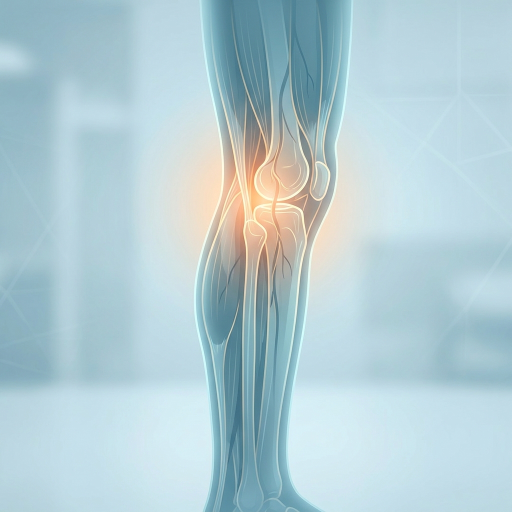
Pain at Back of Knee: The 9-Cause Popliteal Fossa Diagnostic Framework That Maps Every Symptom to Its Source
Introduction: Why Pain at the Back of the Knee Demands a Smarter Approach
A person finishes a morning run and notices a dull ache settling behind the knee. Another individual steps off a long flight and feels a sharp twinge in the same area. A third person simply stands up from a desk chair and experiences unexplained pressure in the popliteal region. Each scenario raises the same question: is this serious, or will it resolve on its own?
Pain at the back of the knee is a symptom description, not a single diagnosis. This discomfort can arise from bones, muscles, tendons, ligaments, bursae, nerves, or blood vessels within the popliteal fossa. Approximately 25% of U.S. adults over age 45 report frequent knee pain, and by age 75, up to 50% experience knee-related discomfort. Yet posterior knee pain remains distinct and often underappreciated compared to anterior, medial, and lateral presentations.
This article introduces a structured, anatomical-layer diagnostic framework: the 9-Cause Popliteal Fossa Framework. This approach maps specific symptom characteristics to their most likely source, helping readers understand which structures may be involved and when to seek medical attention. The framework includes critical red-flag differentiation between conditions such as deep vein thrombosis and ruptured Baker’s cyst, along with severity-tiered treatment pathways ranging from conservative self-care to regenerative medicine options.
This content is educational and designed to support more informed conversations with healthcare providers. It does not replace clinical evaluation.
Anatomy of the Popliteal Fossa: Understanding What Lives Behind the Knee
The popliteal fossa is the diamond-shaped hollow at the back of the knee, bounded by the hamstring muscles superiorly and the gastrocnemius heads inferiorly. Within this compact space lies a complex network of structures that can each become a source of posterior knee pain.
Five anatomical layers generate pain in this region:
- Vascular structures: Popliteal artery and vein
- Ligamentous structures: Posterior cruciate ligament (PCL) and posterior joint capsule
- Musculotendinous structures: Hamstring tendons, popliteus tendon, and gastrocnemius origin
- Bursae: Semimembranosus bursa (Baker’s cyst location)
- Neurological and referred pain sources: Tibial nerve, common peroneal nerve, and lumbar spine referral
This layered anatomy matters because overlapping structures mean that pain location alone is insufficient for diagnosis. Symptom character, onset, aggravating factors, and associated signs must be combined with anatomical knowledge for accurate assessment.
A 2024 review in Insights into Imaging identified a diagnostic gap: current standard ultrasound protocols do not thoroughly investigate the complex posterolateral knee structures, meaning even imaging can miss certain conditions.
The 9-Cause Popliteal Fossa Diagnostic Framework: Mapping Symptoms to Their Source
The framework organizes nine distinct causes by anatomical layer, each with a characteristic symptom profile. Readers should note symptom onset (acute versus gradual), character (aching, sharp, throbbing, burning), location within the popliteal fossa (central, medial, lateral), aggravating activities, and associated signs (swelling, warmth, numbness).
The framework narrows probability, not certainty. Clinical evaluation, physical examination, and imaging remain necessary for definitive diagnosis. Some causes are common and benign; others are rare but potentially life-threatening.
Cause 1: Baker’s Cyst (Popliteal Cyst)
A Baker’s cyst is a fluid-filled sac that forms in the semimembranosus-gastrocnemius bursa when excess synovial fluid accumulates. Prevalence ranges from 5 to 38% in the general population, rising to 20 to 40% in patients with knee osteoarthritis. Most cases occur in adults aged 35 to 70.
Characteristic symptoms: A soft, round lump behind the knee; dull aching or pressure sensation; stiffness worsening with activity or prolonged standing. Many cysts remain asymptomatic.
Baker’s cysts are almost always secondary to an intra-articular disorder such as osteoarthritis, meniscus tear, or rheumatoid arthritis. Treating the cyst without addressing the underlying condition leads to recurrence.
A ruptured Baker’s cyst causes sudden worsening of pain, calf swelling, redness, and warmth. These symptoms closely mimic DVT, a phenomenon called pseudothrombophlebitis syndrome.
In children (peak ages 4 to 7), Baker’s cysts are often idiopathic and frequently resolve without treatment.
Treatment tiers: Conservative management addresses the underlying condition with activity modification and ice. Aspiration with or without corticosteroid injection provides intermediate relief. Surgical excision is reserved for refractory cases. Regenerative options include PRP or hyaluronic acid injection into the joint to address the underlying osteoarthritis driving fluid production.
Cause 2: Posterior Cruciate Ligament (PCL) Injury
The PCL is the primary restraint against posterior tibial translation, running along the back of the knee. It is approximately twice as strong as the ACL, making isolated tears less common. PCL injuries account for less than 20% of all knee ligament injuries, with approximately half resulting from high-energy trauma such as dashboard impact in car accidents.
Characteristic symptoms: Posterior knee pain and instability, often with less dramatic swelling than ACL tears; pain worsening when going downstairs or during deceleration.
Treatment tiers: Grade I and II injuries respond to conservative care including RICE, bracing, and physical therapy. Grade III or combined ligament injuries may require surgical reconstruction. Regenerative options such as PRP or BMAC injection support ligament healing in partial tears, potentially reducing progression to surgical intervention.
Cause 3: Posterior Meniscus Tear
The posterior horn of the medial meniscus is the most common location for meniscus tears because it absorbs the greatest load. More than 40% of people aged 70 and older have experienced a torn meniscus.
Characteristic symptoms: Posterior medial knee pain, joint line tenderness, pain with deep squatting or pivoting, occasional locking or catching sensation, and swelling that develops over hours.
Radial tears of the posterior horn are associated with significantly more severe cartilage degradation than other tear types, making them a key risk factor for progressive knee osteoarthritis.
Treatment tiers: Conservative management with physical therapy and activity modification works well for degenerative tears in older patients. Arthroscopic repair is preferred for younger patients with repairable tears. PRP injection has shown promise in supporting meniscal healing and reducing inflammation.
Cause 4: Hamstring Tendinopathy
Distal hamstring tendinopathy involves degeneration or reactive changes in the semimembranosus, biceps femoris, or semitendinosus tendons near the posterior knee. The semimembranosus tendon is most commonly affected.
Characteristic symptoms: Gradual onset of deep aching pain at the back of the knee, worse with running (especially hills or speed work), cycling, or prolonged sitting with the knee flexed; localized tenderness; no significant swelling.
Treatment tiers: Load management and activity modification come first. Progressive tendon loading rehabilitation follows. PRP injection under ultrasound guidance stimulates collagen remodeling for refractory cases.
Cause 5: Gastrocnemius Strain or Tendinopathy
The gastrocnemius muscle originates from the posterior femoral condyles, placing its proximal attachment within the popliteal fossa.
Two distinct presentations occur: acute gastrocnemius strain (sudden sharp pain during explosive activity, often described as a “pop”) and proximal gastrocnemius tendinopathy (gradual onset with running or stair climbing).
Characteristic symptoms: Pain at the posteromedial or posterolateral knee, calf tightness, pain with ankle dorsiflexion, and tenderness at the gastrocnemius origin.
Treatment tiers: RICE for acute strains; progressive calf strengthening and eccentric loading for tendinopathy; PRP injection at the proximal origin for refractory cases.
Cause 6: Popliteus Tendinopathy or Strain
The popliteus is a small muscle that unlocks the knee from full extension and provides posterolateral rotatory stability. Popliteus tendinopathy is rare but underdiagnosed, particularly in runners and cyclists.
Characteristic symptoms: Lateral or posterolateral knee pain worsening with downhill running, pivoting, or internal rotation of the tibia; tenderness at the lateral femoral condyle; no instability.
Standard ultrasound protocols often fail to adequately visualize the popliteus myotendinous junction, making MRI the preferred diagnostic modality.
Cause 7: Knee Osteoarthritis Affecting the Posterior Compartment
Knee osteoarthritis generates posterior knee pain when degenerative changes affect the posterior compartment or drive Baker’s cyst formation. Globally, knee OA had approximately 374.7 million prevalent cases in 2021, projected to reach approximately 658 million by 2045.
Women consistently show higher prevalence, incidence, and disability than men. Obesity increases knee OA risk by up to four times.
Characteristic symptoms: Diffuse posterior knee aching, morning stiffness lasting less than 30 minutes, pain worsening with activity and improving with rest, crepitus, and reduced range of motion.
Treatment tiers: Weight management, low-impact exercise, and NSAIDs form the foundation. Physical therapy strengthens supporting muscles. Corticosteroid or hyaluronic acid injections manage symptoms. Regenerative options including PRP, BMAC, stem cell therapy, and exosome therapy offer alternatives to surgery. Total knee replacement is indicated only after exhausting appropriate conservative options.
Cause 8: Referred Pain from the Lumbar Spine or Patellofemoral Joint
Pain perceived in the posterior knee can originate from the lumbar spine (L3 to S1 nerve roots) or the patellofemoral joint.
Lumbar spine referral presents with posterior knee pain without a clear local cause, associated with back pain or sciatica, no tenderness on direct popliteal fossa palpation, and neurological symptoms.
Nerve entrapment in the popliteal fossa by a Baker’s cyst, lipoma, or ganglion cyst produces burning, tingling, or electric-shock pain radiating into the calf or foot.
Cause 9: Deep Vein Thrombosis (DVT) of the Popliteal Vein
Popliteal vein DVT is a blood clot forming behind the knee, classified as a proximal DVT with a high risk of pulmonary embolism if untreated. DVT affects approximately 1 in 1,000 people per year, and up to 30 to 40% of cases are asymptomatic.
Characteristic symptoms: Sudden onset of posterior knee pain, calf swelling, warmth, redness, and tenderness along the vein.
Key risk factors: Prolonged immobility, recent surgery, pregnancy, oral contraceptive use, cancer, obesity, and clotting disorders.
DVT can be clinically indistinguishable from a ruptured Baker’s cyst. This is a life-threatening diagnostic dilemma requiring urgent imaging. DVT requires immediate medical evaluation and anticoagulation therapy.
Red Flag Differentiation: When Posterior Knee Pain Requires Immediate Medical Attention
The DVT versus ruptured Baker’s cyst dilemma is critical: both present with sudden posterior knee and calf pain, swelling, warmth, and redness. Ultrasound is the only reliable way to differentiate them.
Seek emergency care immediately for:
- Sudden severe swelling, warmth, and redness behind the knee with calf involvement
- Shortness of breath, chest pain, or rapid heart rate (possible pulmonary embolism)
- Numbness, coldness, or pallor in the foot (possible popliteal artery injury)
- Fever with joint swelling and warmth (possible septic arthritis)
Seek urgent medical evaluation for:
- Inability to bear weight after injury
- Visible deformity or gross instability
- Posterior knee pain following high-energy trauma
- Rapidly worsening pain despite rest and ice
When in doubt, seek medical evaluation rather than self-diagnosing.
Treatment Pathways: From Conservative Care to Regenerative Medicine
Treatment selection depends on specific diagnosis, severity, patient age, activity goals, and response to prior treatment.
Tier 1: Acute and Conservative Self-Care
The RICE method (Rest, Ice, Compression, Elevation) serves as first-line treatment. Ice application for 15 to 20 minutes every 2 to 3 hours in the first 48 to 72 hours reduces inflammation. Over-the-counter NSAIDs address inflammation-driven pain.
Complete rest is rarely beneficial beyond the first 48 to 72 hours. Patients should escalate beyond Tier 1 if symptoms do not improve after 2 to 4 weeks.
Tier 2: Physical Therapy and Supervised Rehabilitation
Physical therapy is the cornerstone for tendinopathies, PCL Grade I and II injuries, post-meniscus repair, and OA-related posterior knee pain.
Progressive loading for tendinopathy follows a sequence: isometric loading, isotonic loading with heavy slow resistance, energy storage and release exercises, and sport-specific loading. Learn more about peptide therapy for tendon repair as a complementary approach to rehabilitation.
Tier 3: Regenerative Medicine and Advanced Interventions
Regenerative medicine bridges conservative care and surgery, offering minimally invasive options for patients who have not responded to Tier 1 and 2 treatments.
PRP (Platelet-Rich Plasma): Derived from the patient’s own blood, concentrated growth factors stimulate tissue healing. Applications include Baker’s cyst (treating underlying OA), hamstring tendinopathy, PCL partial tears, and knee OA.
BMAC (Bone Marrow Aspiration Concentrate): Concentrated bone marrow cells address more advanced OA and significant meniscal pathology.
Stem cell therapy: Promotes tissue repair and regeneration. Currently, 224 clinical trials are investigating stem cell therapies for osteoarthritis, with a major $140 million Phase III trial announced in January 2026.
Exosome therapy: Extracellular vesicles facilitate cellular communication and modulate inflammation. Read more about exosome therapy for tendon and cartilage healing.
Hyaluronic acid: Provides joint lubrication and pain relief for OA-driven posterior knee pain.
At Unicorn Bioscience, all injections are administered under ultrasound or X-ray guidance for precision delivery. This approach is particularly important for posterior knee structures given the complex anatomy. More than 90% of stem cell patients at Unicorn Bioscience have not gone on to knee replacement surgery.
As of 2026, the FDA has not approved stem cell, PRP, or exosome products specifically for orthopedic conditions, but substantial clinical evidence supports safety and efficacy when administered by qualified providers within FDA regulatory frameworks.
Tier 4: Surgical Intervention
Surgery is not the first option. Studies suggest up to 80% of patients told they need total knee replacement may not actually require surgery. Explore regenerative medicine alternatives to knee replacement before committing to an invasive procedure.
Surgical options by condition include: Baker’s cyst excision for refractory cases, PCL Grade III ligament reconstruction, arthroscopic meniscus repair or partial meniscectomy, and surgical decompression for popliteal artery entrapment syndrome.
Prevention: Reducing the Risk of Posterior Knee Pain
Modifiable risk factors include obesity, sedentary lifestyle, poor running mechanics, inadequate warm-up, and prolonged immobility.
Evidence-based prevention strategies include:
- Progressive training load management for athletes
- Hip and glute strengthening to reduce posterior knee load
- Hamstring flexibility and eccentric strength training
- DVT prevention during long travel with hydration, compression stockings, and regular movement
Every pound of body weight adds approximately four pounds of force across the knee joint. Even modest weight loss of 5 to 10% significantly reduces knee pain and OA progression.
Conclusion: From Symptom to Source
Pain at the back of the knee is not a single condition but a symptom arising from nine distinct causes across five anatomical layers of the popliteal fossa. By systematically considering symptom onset, character, location, aggravating factors, and associated signs, clinicians and patients can meaningfully narrow the likely cause and seek appropriate care more efficiently.
DVT and popliteal artery entrapment are medical emergencies requiring immediate evaluation. Most other causes respond to a structured, tiered treatment approach. Surgery is rarely the first or only answer.
With 224 clinical trials investigating stem cell therapy for OA and a major Phase III trial underway in 2026, the regenerative medicine landscape is rapidly expanding. Patients have more options than ever before.
Take the Next Step: Consult with Unicorn Bioscience’s Regenerative Medicine Specialists
For those who have identified a likely cause and are ready to explore treatment options beyond conventional care, Unicorn Bioscience offers expertise in knee conditions including OA, tendinopathy, ligament injuries, and Baker’s cyst secondary to OA. Multiple treatment modalities (PRP, BMAC, stem cell therapy, exosomes, hyaluronic acid) are tailored to individual patient factors.
All injections are administered under ultrasound or X-ray guidance. Same-day treatment is available for qualified candidates, and virtual consultations provide initial assessment options.
Unicorn Bioscience operates 8 locations across Texas (Austin, Dallas, El Paso, Fort Worth, Houston, San Antonio), Florida (Boca Raton), and New York (Manhattan).
Schedule a consultation today by calling (737) 347-0446 or visiting unicornbioscience.com to book a virtual or in-person appointment.
Regenerative medicine treatments are not FDA-approved specifically for orthopedic conditions as of 2026 but are administered within FDA regulatory frameworks by qualified providers. Individual results vary. This article is for educational purposes and does not constitute medical advice.
Schedule Your Consultation Today!
