
Knee on Knee Pain: The Anatomy-First Guide That Explains What’s Actually Happening Inside Your Joint — and What to Do Before Assuming Surgery Is Your Only Option in 2026
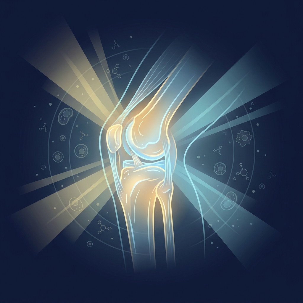
Knee on Knee Pain: The Anatomy-First Guide That Explains What’s Actually Happening Inside Your Joint and What to Do Before Assuming Surgery Is Your Only Option in 2026
Introduction: When Your Knee Feels Like Bone Grinding on Bone
If someone has typed “knee on knee pain” into a search bar, they already know exactly what it feels like. That deep, grinding sensation, the sense that something inside the joint is scraping metal-on-metal with every step, is one of the most distinctive and distressing experiences in all of joint disease. The phrase itself is not a clinical term, but it captures the reality of advanced knee arthritis with remarkable accuracy.
This kind of pain is extraordinarily common. Roughly 25% of adults over the age of 45 in the United States report frequent knee pain, and it ranks among the leading causes of disability in middle-aged and older adults. Those experiencing this condition are far from alone.
This guide makes the following promise: before accepting surgery as inevitable, there are critical anatomical truths and a structured treatment hierarchy that most patients never hear during their first orthopedic consultation. Two revelations, in particular, tend to reframe everything. First, cartilage has no pain fibers, which means the pain is not coming from where most people think it is. Second, an X-ray-based diagnosis may be less severe than a patient has been told.
This article is clinically grounded, honest about limitations, and built to help readers make informed decisions. It is not designed to push anyone toward a single outcome.
What “Knee on Knee Pain” Actually Means: Translating the Body’s Signal
“Knee on knee pain” is a colloquial but anatomically intuitive description of what happens when the cartilage cushioning the joint wears away, allowing the femur (thighbone) and tibia (shinbone) to make direct or near-direct contact. The formal medical term for this condition is knee osteoarthritis (KOA), the most common form of arthritis on the planet.
The scale is staggering. As of 2021, an estimated 374.7 million people worldwide were living with knee osteoarthritis, and knee OA accounts for more than 56% of all osteoarthritis cases globally, according to research published in Arthritis Research & Therapy. This is not a rare or unusual condition. Nearly half of all American adults will develop knee osteoarthritis in at least one knee during their lifetime.
The knee joint is cushioned by articular cartilage covering the ends of the femur, tibia, and patella (kneecap), along with two crescent-shaped shock absorbers called the menisci. The progressive loss of this cushioning tissue defines osteoarthritis. Using everyday language to describe the pain is completely normal, and this guide is designed to bridge that self-description to the clinical framework that actually determines a patient’s treatment options.
The Anatomy of the Knee: A Map to Understanding Pain
To understand where knee pain truly originates, it helps to understand the structures involved:
- Bones: The femur, tibia, patella, and fibula form the joint’s framework.
- Cartilage: Articular cartilage coats the bone ends, and the menisci provide additional cushioning.
- Synovial membrane and fluid: A thin lining that produces lubricating fluid, allowing smooth, frictionless movement.
- Ligaments and tendons: Connective tissues that stabilize the joint and connect muscle to bone.
- Surrounding muscles: The quadriceps, hamstrings, and glutes that support and move the knee.
Articular cartilage is a smooth, shock-absorbing tissue that allows the bones to glide against one another without friction. As it degrades, that gliding surface roughens and thins.
The synovial membrane produces the fluid that lubricates the joint. When it becomes inflamed, a condition called synovitis, it releases pain-signaling chemicals into the joint.
Beneath the cartilage lies the subchondral bone. As cartilage thins, this bone becomes exposed and stressed, making it a major source of pain. Deeper still, bone marrow lesions (BMLs) can develop. These are abnormalities in the subchondral bone that are visible on MRI but invisible on X-ray. They are strongly associated with knee pain and disease progression, and they are frequently overlooked in standard diagnoses.
The Revelation Most Doctors Don’t Explain: Cartilage Cannot Feel Pain
Here is the anatomical truth that changes everything: articular cartilage contains zero pain fibers. It is aneural, meaning it is physically incapable of generating pain signals.
The implication is profound. The agonizing sensation patients describe as “bone on bone” is not coming from the cartilage itself. It originates from the surrounding structures. Specifically, knee OA pain arises from:
- Subchondral bone stress and microfractures as exposed bone absorbs impact it was never designed to handle.
- Synovitis, the inflammation of the joint lining that releases pain-signaling chemicals.
- Bone marrow lesions that create internal pressure within the bone.
- Muscle and tendon strain caused by altered gait and compensating movement patterns.
This explains a critical clinical reality: two patients with nearly identical X-rays can have completely different levels of pain. Imaging severity does not equal pain severity. Research shows that more than half of people who display signs of knee OA on imaging report no pain at all, a disconnect documented across clinical literature.
This revelation is deeply empowering. If pain is coming from the bone, synovium, and soft tissue rather than the cartilage, then treatments targeting those structures can provide genuine relief without removing or replacing the joint.
Why an X-Ray May Be Telling an Incomplete Story
Most patients are never told a fundamental limitation of X-rays: they cannot directly visualize cartilage. X-rays show bone, not soft tissue.
What appears as “joint space narrowing” on an X-ray is only an indirect inference of cartilage loss, based on the gap between the bones. It is not a direct measurement of how much cartilage actually remains.
MRI is the gold standard for direct cartilage assessment, yet many patients receive a “bone-on-bone” label based solely on an X-ray, without ever undergoing an MRI. This imaging gap means “bone-on-bone” diagnoses can be imprecise. A significant number of patients told they are Grade 4 are actually Grade 3, where meaningful cartilage still remains.
Even at Grade 4, research from the MOST Study found ongoing disease activity, including fluctuating bone marrow lesions, joint effusion, and synovitis over a 30-month period. Even “end-stage” knees are not static, and they remain responsive to intervention.
This is not about dismissing a diagnosis. It is about obtaining an accurate one, because the distinction between Grade 3 and Grade 4 dramatically changes which non-surgical treatments are appropriate and likely to succeed.
The Kellgren-Lawrence Grading System: The Knee’s Report Card
The Kellgren-Lawrence (KL) grading system is the standard clinical framework for classifying knee OA severity on X-ray, using a scale from 0 to 4. Understanding a KL grade matters because it is the primary clinical variable that determines which non-surgical treatments carry the strongest evidence for a given stage.
Grade 0: Normal — No Signs of Osteoarthritis
No radiographic features of OA are present. Joint space is normal and no osteophytes (bone spurs) are visible. Pain at this stage typically comes from other causes such as meniscal tears, ligament injuries, bursitis, or referred pain, and requires different evaluation.
Grade 1: Doubtful — Early Warning Signs
Possible minor osteophyte formation, with joint space appearing normal or near-normal. This stage is often asymptomatic or produces mild, intermittent discomfort. Lifestyle interventions such as exercise, weight management, and physical therapy are highly effective here and can slow or halt progression.
Grade 2: Mild — Cartilage Thinning Begins
Definite osteophytes with possible joint space narrowing indicate that cartilage thinning is beginning. Typical symptoms include aching after activity, morning stiffness, and occasional swelling. This is a critical intervention window, where physical therapy, PRP, and hyaluronic acid injections show strong evidence.
Grade 3: Moderate — Significant Cartilage Loss (Often Mislabeled “Bone-on-Bone”)
Multiple osteophytes, definite joint space narrowing, some sclerosis (bone hardening), and possible bone deformity characterize this grade. The crucial nuance is this: Grade 3 means significant cartilage loss but not complete absence. Cartilage remains, and that distinction matters enormously. Many patients told they are “bone-on-bone” are actually Grade 3, and Grade 3 patients are generally good candidates for regenerative and non-surgical interventions. Symptoms tend to be more persistent, including pain during activity and at rest, reduced range of motion, and joint instability.
Grade 4: Severe — True “Bone-on-Bone” Classification
Large osteophytes, marked joint space narrowing, severe sclerosis, and definite bone deformity define this grade. This is the true radiographic definition of “bone-on-bone.” In the interest of transparency, Grade 4 patients are generally poor candidates for stem cell therapy, because regenerative treatments cannot rebuild lost bone structure or severely degraded cartilage. However, even Grade 4 patients may benefit from pain-management interventions such as radiofrequency ablation or genicular artery embolization, which do not aim to regenerate cartilage but can significantly reduce pain and improve function. An accurate grade determination, ideally confirmed with MRI, is essential before accepting surgery as the only path forward.
Mapping Symptoms to Grade: A Self-Assessment Framework
While only imaging interpreted by a clinician can confirm a KL grade, patients can often contextualize their experience using general symptom patterns:
- Early (Grades 1–2): Mild aching after activity, occasional stiffness, and intermittent swelling.
- Moderate (Grade 3): Pain during and after activity, stiffness, swelling, reduced range of motion, and a sense of instability.
- Severe (Grade 4): Constant pain including at rest, significant functional limitation, and visible joint deformity.
Self-assessment is a starting point, not a diagnosis. An accurate KL grade requires imaging interpreted by a qualified clinician. It is also worth considering functional assessment: how the knee actually performs in daily life is as clinically relevant as what the X-ray shows. The goal of this framework is to help patients arrive at their next clinical conversation better informed, knowing what questions to ask and what information to request.
The Non-Surgical Treatment Hierarchy: What to Try Before Surgery
The following is a structured, evidence-based decision pathway, not a random list of alternatives. It moves from foundational interventions to advanced regenerative options. This hierarchy is not about delaying necessary care. It is about ensuring that every appropriate non-surgical option has been genuinely explored before a decision as significant as joint replacement is made. The right starting point depends on a patient’s KL grade, age, activity level, inflammation status, and personal health goals.
Step 1: Physical Therapy and Therapeutic Exercise
Exercise is the foundational intervention for knee OA at all grades. Strengthening the muscles around the knee, including the quadriceps, hamstrings, and glutes, reduces the mechanical load on the joint itself. Physical therapy is not simply “working through pain.” It involves targeted neuromuscular retraining, gait correction, and progressive loading protocols. Exercise therapy is consistently recommended as first-line treatment across clinical guidelines, as noted by Johns Hopkins Medicine. Patients in significant pain often resist exercise, which is exactly why a structured, supervised program with appropriate pain management support is so important.
Step 2: Weight Management
Body weight has a powerful mechanical impact on the knee. Each pound of body weight translates to roughly four pounds of force on the joint during walking. Beyond mechanics, obesity is an independent risk factor for OA because excess adipose tissue secretes inflammatory cytokines that directly accelerate cartilage degradation. Weight management is not a judgment; it is one of the highest-leverage interventions available, and even modest weight reduction produces measurable reductions in knee pain and functional improvement.
Step 3: Unloader Bracing
Unloader braces apply a corrective force that shifts load away from the most damaged compartment of the knee, typically the inner (medial) compartment. Bracing is most effective for unicompartmental OA, where damage is concentrated on one side of the joint, and less effective for severe bilateral disease. It is a practical, non-invasive adjunct that can reduce pain and improve function while other treatments are initiated.
Step 4: Hyaluronic Acid (Viscosupplementation) Injections
Hyaluronic acid (HA) is a naturally occurring component of synovial fluid that provides lubrication and shock absorption. In OA, both its concentration and molecular weight decline. Viscosupplementation involves injecting HA directly into the joint to restore the viscoelastic properties of that fluid, reducing friction and pain. HA injections are most effective for mild to moderate OA (KL Grades 1–3) and can provide meaningful relief for several months. Unicorn Bioscience offers precision-guided HA injections as part of its treatment menu.
Step 5: PRP (Platelet-Rich Plasma) Injections
PRP is derived from the patient’s own blood, concentrated to increase platelet levels, and injected into the joint to deliver growth factors that promote tissue repair and reduce inflammation. A 2025 comprehensive narrative review of 40 high-quality studies, published via the National Library of Medicine, found that leukocyte-poor PRP demonstrates superior pain relief and functional improvement compared to hyaluronic acid and corticosteroids, particularly in KL Grades 1–3. A 2025 meta-analysis in the American Journal of Sports Medicine similarly found PRP provides clinically relevant functional improvement at 1, 3, 6, and 12 months versus placebo, with high-platelet formulations delivering more durable results. Because PRP is autologous (derived from the patient’s own body), immunological risk is minimal. Unicorn Bioscience administers PRP through a precision-guided injection protocol to ensure accurate intra-articular delivery.
Step 6: Stem Cell Therapy (MSC/BMAC)
Mesenchymal stem cell (MSC) therapy involves harvesting stem cells from the patient’s adipose tissue or bone marrow, concentrating them, and injecting them into the joint to promote tissue repair, modulate inflammation, and potentially support cartilage health. A 2025 meta-analysis of 8 randomized controlled trials (502 patients) published in Stem Cell Research & Therapy confirmed that intra-articular MSC therapy significantly reduces pain and improves function for at least 12 months. Notably, adipose-derived MSCs showed better efficacy than bone marrow MSCs, and high-dose treatments produced significantly improved WOMAC scores at six months.
Transparency matters here: stem cell therapy works best for KL Grades 1–3. Patients with severe Grade 4 OA are generally poor candidates, because stem cells cannot regenerate lost bone structure or severely degraded cartilage. As of 2026, the FDA has not approved stem cell therapies for orthopedic conditions, including knee OA, though substantial clinical evidence supports safety and efficacy when administered by qualified providers within FDA regulatory frameworks. Unicorn Bioscience’s BMAC offering is guided by a personalized treatment planning approach that considers patient age, inflammation levels, injury type, and health goals.
Step 7: Exosome Therapy
Exosomes are extracellular vesicles secreted by stem cells that carry signaling molecules such as proteins and RNA, capable of modulating cellular behavior and reducing inflammation. In knee OA, exosomes may facilitate cellular communication that promotes tissue repair and suppresses inflammatory pathways without introducing live cells. This is an emerging modality with growing clinical interest, and Unicorn Bioscience offers it within its multi-modal treatment menu. As with all regenerative therapies, it should be received from qualified providers operating within appropriate regulatory frameworks.
Step 8: Radiofrequency Ablation (RFA) for Knee Pain
Genicular nerve RFA is a minimally invasive procedure that uses radiofrequency energy to disrupt the pain signals transmitted by the genicular nerves, which carry knee pain signals to the brain. RFA does not treat the underlying OA, but it provides significant and durable pain relief. This makes it valuable for patients who are not candidates for regenerative therapy (including those with severe Grade 4 OA) or who need pain control while pursuing other interventions. Performed under imaging guidance as an outpatient procedure with minimal recovery time, RFA is particularly useful for patients whose pain is limiting their ability to engage in physical therapy.
Step 9: Genicular Artery Embolization (GAE)
GAE is an emerging, minimally invasive vascular intervention. This catheter-based procedure reduces blood flow to the inflamed synovial tissue of the knee, decreasing the inflammatory environment that drives pain. GAE shows a 99.7% technical success rate with pain reduction of 34 to 39 points on the Visual Analog Scale. The mechanism targets abnormal neovascularization (new blood vessel growth) in the synovium, a known driver of OA pain. GAE is particularly valuable for patients with significant synovitis-driven pain who have not responded adequately to injection therapies, and it is covered in a 2026 peer-reviewed perspective on surgical and nonsurgical treatment of knee osteoarthritis published in the National Library of Medicine.
A Note on Corticosteroid Injections: Why They Are Not a Long-Term Strategy
Corticosteroid (cortisone) injections are widely used and can provide meaningful short-term pain relief, typically lasting weeks to a few months. However, an important clinical caveat is rarely explained: studies show that repeated corticosteroid injections may accelerate cartilage breakdown over time, potentially worsening the very condition they are meant to manage.
The practical implication is that corticosteroids are best used as a short-term bridge, reducing acute inflammation to allow participation in physical therapy, rather than as a primary or repeated long-term treatment. This contrasts with regenerative approaches like PRP and MSC therapy, which aim to address the biological drivers of OA rather than simply suppressing symptoms. Patients often receive repeated cortisone injections without being informed of these long-term trade-offs.
The Surgery Question: When Is Total Knee Replacement Actually Necessary?
Total knee arthroplasty (TKA) is a legitimate and effective procedure for the right patient. This guide is not anti-surgery; it is pro-informed decision-making.
The scale is significant. Annual primary TKA volume rose from about 603,704 procedures in 2013 to over 1 million by 2022, and is projected to reach nearly 2.8 million by 2040, according to data published in PubMed. Yet research suggests that up to 80% of patients told they need total knee replacement may not actually require surgery, which underscores the value of thorough non-surgical evaluation.
Appropriate TKA candidates typically have confirmed KL Grade 4 OA, severe functional limitation, failure of multiple non-surgical interventions, and significant quality-of-life impairment. TKA is a major surgical procedure with a considerable recovery period, potential complications, and a finite implant lifespan, factors that carry particular weight for younger or more active patients. For a detailed overview of what this entails, reviewing knee replacement surgery risks and complications can help patients weigh their options. Surgery should follow a genuine trial of appropriate non-surgical interventions, not precede it.
The Psychological Dimension: Why “Bone-on-Bone” Feels Like a Death Sentence for the Knee
A “bone-on-bone” diagnosis carries real emotional weight. For many patients, it triggers fear, fatalism, and a sense that surgery is inevitable, often before they have explored the full range of options. The language itself contributes to this response. “Bone-on-bone” is viscerally alarming, conjuring an image of irreversible mechanical destruction.
The anatomical truths established earlier reframe the picture: the pain comes from treatable structures (bone, synovium, and soft tissue); the X-ray may be overstating severity; and the KL grade rather than the colloquial label determines treatment candidacy. This matters because catastrophizing is well documented in chronic pain. Patients who believe their condition is worse than it is tend to experience more pain and worse outcomes, which makes accurate information a therapeutic intervention in its own right. Approaching the next clinical conversation as an informed participant rather than a passive recipient changes the entire trajectory.
Who Is a Good Candidate for Non-Surgical and Regenerative Treatment?
Candidacy is best understood through KL grade, patient profile, and treatment goals:
- Ideal candidates for regenerative therapy (PRP, MSC/stem cell): KL Grades 1–3, patients with meaningful cartilage remaining, those motivated to engage in complementary rehabilitation, and individuals seeking to delay or avoid surgery.
- Candidates for pain-management interventions (RFA, GAE) regardless of grade: Patients with severe pain limiting function, those who are not surgical candidates for medical reasons, and those seeking relief while pursuing other treatments.
- Candidates for whom surgery may be most appropriate: Confirmed KL Grade 4 with complete cartilage loss, severe functional limitation, failure of multiple well-executed non-surgical interventions, and significant quality-of-life impairment.
Candidacy determination requires personalized assessment, including imaging review, functional evaluation, and discussion of health goals. Unicorn Bioscience develops treatment protocols based on individual factors including inflammation levels, patient age, injury type, current medications, and personal health goals.
Why Accurate Diagnosis Matters More Than the Label Given
The core argument of this guide is straightforward: the colloquial label “bone-on-bone” matters far less than knowing an actual KL grade, understanding where the pain originates, and having a treatment plan calibrated to specific anatomy and goals.
If a “bone-on-bone” diagnosis was based solely on X-ray, an MRI may reveal more cartilage than the X-ray suggested and a different KL grade that opens additional treatment options. This is why seeking a second opinion or a specialized regenerative medicine evaluation before committing to surgery is so valuable, especially for patients who have not been offered a structured non-surgical pathway. Unicorn Bioscience offers both virtual and in-person consultations across Texas, Florida, and New York, making an expert evaluation accessible without significant logistical burden. The evaluation is an information-gathering step, not a commitment.
Conclusion: The Knee Has More Options Than Most Patients Are Told
Four revelations should stay with anyone experiencing knee on knee pain. First, cartilage cannot feel pain; the pain comes from bone, synovium, and soft tissue, all of which are treatable. Second, X-rays cannot directly visualize cartilage, so “bone-on-bone” diagnoses can be imprecise. Third, many patients labeled KL Grade 4 are actually Grade 3, where non-surgical options carry stronger evidence. Fourth, a structured, evidence-based treatment hierarchy exists between “do nothing” and “replace the joint.”
Knee OA affects hundreds of millions of people worldwide, and the trajectory is worsening. Yet the non-surgical treatment landscape is advancing rapidly, with significant clinical trial investment and growing evidence for regenerative approaches. Not every patient is a candidate for every treatment, and some will ultimately benefit most from surgery. But that determination should follow a thorough, personalized evaluation, not arrive as a first response to an alarming label. Understanding one’s anatomy, KL grade, and treatment options transforms a passive patient into an informed decision-maker, and that knowledge is the most important first step.
Ready to Find Out Where You Actually Stand? Start With a Personalized Evaluation
The next concrete step is a consultation with Unicorn Bioscience to determine an actual KL grade, identify the specific sources of pain, and explore which non-surgical or regenerative treatments are appropriate for a given situation.
Unicorn Bioscience offers a multi-modal treatment menu including PRP, stem cell and BMAC therapy, exosomes, hyaluronic acid, and peptide therapy, all delivered through precision imaging-guided injections and personalized treatment protocols, with same-day treatment availability for qualified candidates. Consultations, both virtual and in-person, are available across 8 locations in Texas (Austin, Dallas, El Paso, Fort Worth, Houston, and San Antonio), Florida (Boca Raton), and New York (Manhattan).
A consultation is an information-gathering step. The goal is to provide the clinical clarity needed to make the best decision for a knee and a life. To schedule a virtual or in-person consultation, contact Unicorn Bioscience at (737) 347-0446 or visit unicornbioscience.com.
Schedule Your Consultation Today!
