
Aching Pain Behind Knee: The Symptom-Character Diagnostic Framework That Separates Baker’s Cyst, Semimembranosus Tendinopathy, and Early OA
Aching Pain Behind Knee: The Symptom-Character Diagnostic Framework That Separates Baker’s Cyst, Semimembranosus Tendinopathy, and Early OA
Introduction: Why ‘Aching’ Behind the Knee Is a Diagnostic Clue, Not Just a Symptom
A persistent, dull ache behind the knee that refuses to fade represents more than a minor inconvenience. Unlike sharp, stabbing, or burning pain, this specific quality of discomfort carries meaningful diagnostic information. The character of the ache itself serves as a clinical signal that can help narrow down the underlying cause before any imaging is performed.
Whether patients describe it as “aching pain behind knee,” “ache pain behind knee,” or “achy pain behind knee,” they are describing the same symptom cluster. This article addresses all three variations together, providing a comprehensive framework for understanding what this particular type of discomfort may indicate.
Three conditions most commonly produce this dull, persistent posterior knee aching: Baker’s cyst (popliteal cyst), semimembranosus tendinopathy, and early posterior knee osteoarthritis. While these conditions share similar symptom profiles, they require different treatment approaches. Misidentifying the source of the aching can lead to months of ineffective treatment.
This article provides a symptom-character triage framework that helps differentiate these three conditions. It also addresses two critical safety considerations that most patient-facing content overlooks: the deep vein thrombosis (DVT) emergency that can mimic a ruptured Baker’s cyst, and the kinetic chain factors upstream from the knee that often contribute to persistent posterior knee pain.
The stakes are significant. Knee osteoarthritis alone affects approximately 528 million people worldwide. Contrary to common assumptions, more than half of individuals with symptomatic knee OA are younger than 65. Posterior knee aching is not exclusively a problem of the elderly.
This article is educational and informational. It does not replace clinical evaluation. Certain symptom combinations require same-day medical care, and these will be flagged clearly throughout.
Anatomy of the Posterior Knee: Why So Many Conditions Produce the Same Aching Sensation
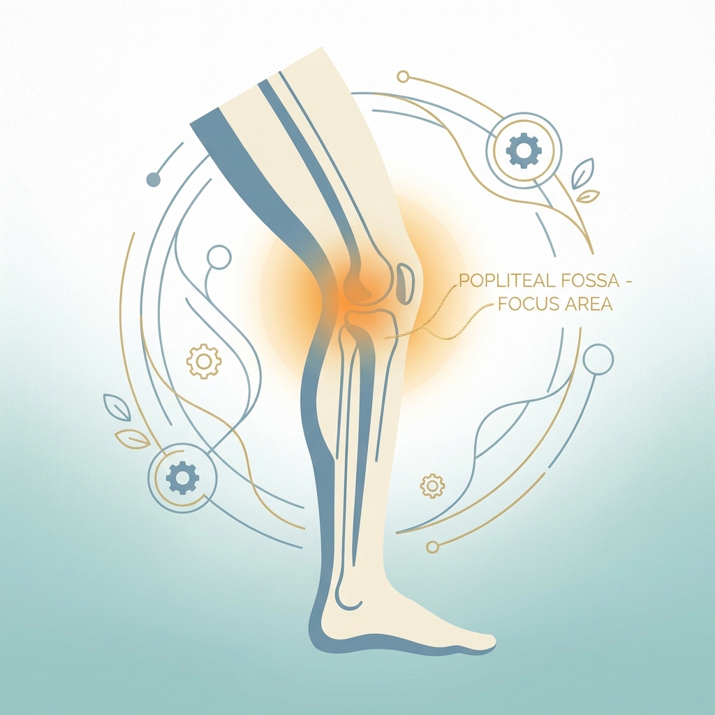
The popliteal fossa, the diamond-shaped space behind the knee, is a dense anatomical crossroads rather than a simple hollow. This region contains the semimembranosus, semitendinosus, and biceps femoris tendons of the hamstring group. It also houses the gastrocnemius muscle origin, the popliteus muscle, the popliteal bursa, major nerves, and critical blood vessels including the popliteal artery and vein.
This anatomical density explains why multiple distinct conditions can produce remarkably similar aching sensations. A fluid-filled cyst, a degenerating tendon, early cartilage loss, or even a blood clot can all generate a dull, persistent ache in the same general region.
Referred pain adds another layer of complexity. The lumbar spine, particularly lumbosacral radiculopathy involving the L4-S1 nerve roots, can produce aching that the brain perceives as originating behind the knee. The patellofemoral joint can also generate referred posterior knee discomfort. These frequently missed diagnostic angles can lead to months of mismanaged local knee treatment when the actual source lies elsewhere.
Because the anatomy is complex, the quality, location, and behavior of the ache become the primary tools for initial triage. Understanding these distinctions helps patients communicate more precisely with their clinicians and ask the right questions.
The Symptom-Character Diagnostic Framework: Using the Quality of the Ache to Narrow the Cause
The specific character of aching pain provides meaningful diagnostic signal. Its location, behavior, triggers, and accompanying sensations all contribute to identifying the likely source.
A foundational distinction exists between chronic and acute presentations. Dull, constant aching that builds gradually is more characteristic of chronic conditions such as tendinopathy, osteoarthritis, or an intact Baker’s cyst. Sharp pain that worsens with specific movements suggests acute mechanical injury, including meniscus tears or a ruptured cyst.
The three-condition comparison that follows serves as a practical triage tool rather than a definitive self-diagnosis. The goal is to help patients understand their symptoms more precisely and prepare for productive conversations with their healthcare providers.
Condition 1: Baker’s Cyst (Popliteal Cyst): Central Fullness and Pressure-Ache
A Baker’s cyst is a fluid-filled sac that forms in the popliteal fossa between the semimembranosus tendon and the medial head of the gastrocnemius muscle. It develops when excess synovial fluid from underlying joint pathology, most commonly osteoarthritis or meniscal tears in adults, accumulates in this space.
Baker’s cysts affect approximately 20% of the general population. Among patients with knee pain specifically, prevalence reaches 25.8% based on ultrasonographic studies. In knee osteoarthritis patients, the prevalence ranges from 20% to 40%. Higher BMI and heavy occupational work are significant risk factors in patients with both OA and Baker’s cyst.
The characteristic aching pattern involves central posterior fullness and a pressure-ache rather than sharp pain. Patients often describe it as “tightness” or a sensation of “something behind the knee.” The discomfort is most noticeable when standing after prolonged sitting or during full knee extension.
Importantly, the ache from a Baker’s cyst is secondary. The cyst itself is a symptom of underlying joint pathology, not the primary disease. Treating the underlying cause, whether osteoarthritis or a meniscal tear, typically resolves the cyst.
On self-examination, a Baker’s cyst may feel like a soft, fluctuant bulge in the popliteal fossa. It is sometimes visible when the knee is extended and may be painless or mildly tender.
The key distinction from semimembranosus tendinopathy is location: Baker’s cyst produces central fullness and diffuse pressure, while tendinopathy produces a pinpoint posteromedial ache inferior to the joint line.
Treatment for most Baker’s cysts involves addressing the underlying cause. For large, painful, or repeatedly rupturing cysts, aspiration combined with corticosteroid injection is the standard non-surgical intervention. Surgical excision is rarely needed.
The Ruptured Baker’s Cyst: A Same-Day Emergency That Mimics DVT
New or sudden calf swelling combined with posterior knee pain is a same-day medical emergency until DVT is ruled out.
Popliteal vein thrombosis can itself present as a dull ache behind the knee. Approximately 50% of DVT cases produce no noticeable symptoms, making awareness of risk factors essential. These include prolonged immobility, recent surgery, obesity, pregnancy, clotting disorders, and long-haul travel.
Doppler ultrasound is the most reliable method to distinguish a ruptured Baker’s cyst from DVT. Both conditions can be assessed in the same imaging session.
Patients should not wait to see if calf swelling resolves on its own. DVT can lead to pulmonary embolism, which is life-threatening.
Condition 2: Semimembranosus Tendinopathy: The Underdiagnosed Cause of Posteromedial Knee Aching
Semimembranosus tendinopathy represents a clinically underdiagnosed cause of chronic posteromedial knee pain. It is rarely described in the medical literature and may be inadequately treated due to lack of clinical awareness. This represents a major gap in most patient-facing content.
The semimembranosus is one of three hamstring muscles. Its distal tendon inserts at the posteromedial aspect of the tibia, placing it directly in the region patients describe as “behind and slightly inside the knee.”
The characteristic aching pattern involves an insidious, progressive ache at the posteromedial knee. Tenderness on palpation inferior to the joint line distinguishes it from Baker’s cyst, which produces central fullness. The pain worsens with running, cycling, walking downstairs, or sudden deep knee flexion.
Semimembranosus tendinopathy is commonly misdiagnosed as Baker’s cyst or medial meniscal pathology due to anatomical proximity and similar aching quality.
The patient demographic most likely to present with this condition includes active adults, runners, cyclists, and individuals who perform repetitive knee flexion-extension activities. These patients are often younger than the typical OA patient. For athletes dealing with this type of chronic tendon degeneration, cellular therapy for athletes represents an emerging treatment consideration.
The tendinopathy mechanism involves repetitive loading without adequate recovery, leading to tendon degeneration (tendinosis) rather than acute inflammation. This is why rest alone is insufficient and may worsen deconditioning.
Treatment centers on education and progressive loading exercise targeting kinetic chain function and hamstring loading capacity. A 2025 randomized controlled trial involving 100 participants found no significant difference between individualized physiotherapy and shockwave therapy at 12 months. Aggressive stretching alone is not recommended and may worsen symptoms. For patients seeking regenerative options, hamstring tendonitis cellular therapy offers a targeted non-surgical approach to tendon healing.
MRI can confirm tendon degeneration, while ultrasound can identify tendon thickening and peritendinous changes.
Condition 3: Early Posterior Knee Osteoarthritis: The Diffuse Ache That Builds Over Time
Osteoarthritis can produce posterior knee aching through multiple mechanisms: posterior compartment cartilage loss, synovial inflammation, osteophyte formation, and secondary Baker’s cyst development.
The global burden is substantial. Knee OA affects approximately 528 million people worldwide. Global prevalence is 16.0% in adults aged 15 and over and 22.9% in those aged 40 and over. Prevalence has doubled in the United States since the mid-20th century, independent of age and BMI alone.
The age misconception deserves emphasis: more than half of individuals with symptomatic knee OA are younger than 65. Early posterior OA is not exclusively a condition of the elderly.
The characteristic aching pattern of early posterior OA involves diffuse, deep aching that is worse after periods of inactivity, particularly morning stiffness. It worsens with prolonged weight-bearing and may be accompanied by mild swelling or warmth. Unlike semimembranosus tendinopathy, the ache is not localized to a single tendon insertion point.
The OA-Baker’s cyst connection is important: OA is the most common underlying cause of Baker’s cyst in adults. Patients with both conditions experience a combined pressure-ache and diffuse joint aching.
Current evidence from 2025 and 2026 supports a layered multimodal approach to treatment. Conservative care comes first, including exercise, weight management, and NSAIDs. Injections follow, including corticosteroids, hyaluronic acid, and PRP. Neuromodulation options include genicular nerve radiofrequency ablation and COOLIEF cooled RFA, which provides up to 24 months of pain relief. Genicular artery embolization represents another option before considering surgery.
Emerging treatments include investigational iPSC-derived nociceptor therapy and a major Phase III clinical trial announced in January 2026. The field is evolving rapidly.
Side-by-Side Comparison: How to Tell the Three Conditions Apart
| Dimension | Baker’s Cyst | Semimembranosus Tendinopathy | Early Posterior OA |
|---|---|---|---|
| Pain Location | Central posterior fullness | Posteromedial pinpoint | Diffuse posterior and medial |
| Pain Quality | Pressure, fullness, tightness | Localized tendon ache | Deep joint ache |
| Primary Triggers | Full extension, prolonged standing | Hamstring-loading activities | Morning stiffness, prolonged weight-bearing |
| Accompanying Signs | Palpable bulge | Tendon tenderness inferior to joint line | Mild swelling, crepitus |
| Typical Patient | Adults 35-70 with OA or meniscal history | Active adults, runners | Adults 40+ with gradual onset |
| Response to Rest | Partial relief | Worsens with complete rest | Partial relief with activity modification |
If the aching is accompanied by lower back pain, buttock pain, or radiating symptoms down the leg, lumbar radiculopathy or patellofemoral referred pain should be considered. These warrant clinical evaluation.
This comparison is a communication tool to help patients describe their symptoms more precisely to a clinician. It is not a substitute for professional diagnosis.
The Kinetic Chain Factor: Why the Source of Posterior Knee Aching May Not Be in the Knee
The knee is a middle-link joint. Dysfunction above the knee (hip, lumbar spine) or below it (ankle, foot) alters the mechanical load placed on posterior knee structures.
Hip weakness serves as a primary upstream driver. Weak hip abductors and external rotators cause the femur to internally rotate and adduct during weight-bearing. This increases valgus stress and alters hamstring mechanics, directly increasing load on the semimembranosus tendon and posterior knee structures.
Ankle dysfunction contributes as well. Limited ankle dorsiflexion from tight calves, prior sprains, or Achilles stiffness forces compensatory knee flexion patterns that increase posterior knee compressive and tensile loads. Patients with a history of ankle ligament tears may be particularly susceptible to these downstream mechanical compensations.
The lumbar spine can also contribute. Lumbosacral radiculopathy, particularly L4-S1 nerve root involvement, can produce referred aching perceived as originating behind the knee. This missed diagnosis leads to months of mismanaged local knee treatment. Patients with concurrent back symptoms may benefit from understanding spine regenerative injection therapy as part of a comprehensive treatment approach.
The practical implication is significant: regardless of whether the primary diagnosis is Baker’s cyst, semimembranosus tendinopathy, or early OA, addressing kinetic chain dysfunction is essential for durable recovery. Treating only the local structure without addressing upstream causes leads to recurrence.
Hip strengthening exercises such as clamshells and lateral band walks, ankle mobility work including calf stretching and dorsiflexion drills, and core stability exercises are frequently included in rehabilitation programs for all three posterior knee conditions.
Red Flags: When Aching Pain Behind the Knee Requires Same-Day or Urgent Care
Most aching behind the knee is not a medical emergency. However, certain symptom combinations require immediate evaluation.
Same-day emergency symptoms:
- Sudden calf swelling with posterior knee pain (ruptured Baker’s cyst vs. DVT cannot be distinguished without imaging)
- Calf warmth, redness, or tenderness accompanying posterior knee aching
- Shortness of breath or chest pain following posterior knee or calf symptoms (possible pulmonary embolism)
Urgent symptoms (within 24-48 hours):
- Posterior knee aching accompanied by significant joint swelling, fever, or redness (possible septic arthritis or inflammatory flare)
- Acute locking or giving way of the knee with posterior aching (possible meniscal tear or loose body)
- Posterior knee pain following direct trauma
Symptoms warranting professional evaluation within 1-2 weeks:
- Aching that does not improve within 2-3 weeks of consistent home care
- Aching that wakes the patient from sleep
- Aching accompanied by unexplained weight loss or fatigue
Doppler ultrasound serves as the first-line imaging tool for DVT exclusion. MRI provides soft tissue assessment for meniscus, tendons, and cysts when the clinical picture is unclear.
Conservative Home Care: What Helps and What Can Make It Worse
Conservative treatment is effective for most patients with posterior knee aching from Baker’s cyst, semimembranosus tendinopathy, or early OA. However, the appropriate approach depends on the underlying cause.
General measures applicable to all three conditions:
- Activity modification (reduce aggravating activities without complete rest)
- Ice for acute flares (15-20 minutes, several times daily)
- Over-the-counter NSAIDs for pain and inflammation management
- Elevation for swelling
Baker’s cyst-specific guidance: Treating the underlying cause (OA management, meniscal treatment) is the primary strategy. The cyst itself does not require direct treatment in most cases. Aggressive compression that may rupture the cyst should be avoided.
Semimembranosus tendinopathy-specific guidance: Progressive loading exercise, not rest alone, is the cornerstone of tendinopathy recovery. Aggressive passive stretching of the hamstring can worsen tendon irritation and is not recommended as a standalone treatment. A graded return-to-activity program supervised by a physiotherapist is optimal.
Early OA-specific guidance: Low-impact aerobic exercise (swimming, cycling, walking) maintains joint health. Weight management reduces posterior knee load. NSAIDs provide symptomatic relief. Prolonged immobility, which accelerates deconditioning, should be avoided.
Common mistakes to avoid:
- Complete rest for tendinopathy (worsens deconditioning)
- Aggressive hamstring stretching for nerve-driven or tendon-driven aching (can worsen symptoms)
- Ignoring calf swelling as “just a muscle cramp” (DVT risk)
Pain that does not improve within 2-3 weeks of consistent home care warrants professional evaluation.
Professional Treatment Options: From Injections to Regenerative Medicine
Professional treatment progresses from conservative to more advanced interventions, aligned with 2025-2026 evidence-based guidelines.
Physical therapy forms the foundation of professional treatment for all three conditions. It addresses kinetic chain dysfunction, progressive loading, and movement pattern correction.
Aspiration and corticosteroid injection for Baker’s cyst represents the standard non-surgical intervention for large, painful, or repeatedly rupturing cysts. Ultrasound-guided injection improves accuracy and outcomes. This provides symptomatic relief but does not address the underlying cause.
Injection therapies for OA include corticosteroid injections for acute inflammatory flares, hyaluronic acid knee injection for joint lubrication and pain relief, and PRP (platelet-rich plasma) as an emerging option with a growing evidence base.
For patients with posterior knee aching driven by early OA or tendon degeneration who want to avoid surgery, regenerative medicine approaches represent an evolving treatment frontier. Unicorn Bioscience offers multiple regenerative modalities including PRP, BMAC, stem cell therapy, exosome therapy, and hyaluronic acid injections. All treatments are administered under precision ultrasound or X-ray guidance for accurate delivery to the affected structures.
As of 2026, the FDA has not approved stem cell, PRP, or exosome products specifically for orthopedic conditions. However, substantial clinical evidence supports safety and efficacy when administered by qualified providers within FDA regulatory frameworks. Patients interested in understanding the evidence base for these approaches can review the regenerative medicine clinical evidence framework for a detailed overview.
Advanced interventions for refractory OA pain include genicular nerve radiofrequency ablation, COOLIEF cooled RFA (providing up to 24 months of pain relief), and genicular artery embolization with a 99.7% technical success rate and 34-39 point VAS pain reduction.
Surgical options are reserved for cases where conservative and minimally invasive treatments have failed. Baker’s cyst surgical excision is rarely needed. Knee replacement remains a last resort, with evidence suggesting up to 80% of patients told they need total knee replacement may not actually require surgery.
Accurate diagnosis before treatment is essential. The wrong treatment for the wrong condition will not resolve the aching and may delay appropriate care.
When to See a Doctor: A Practical Decision Guide
Tier 1: Call emergency services or go to the ER immediately
- Sudden calf swelling with posterior knee pain
- Shortness of breath or chest pain following leg symptoms
- Signs of severe infection (fever, extreme redness, hot joint)
Tier 2: Seek same-day urgent care
- New calf swelling or warmth with posterior knee aching (DVT must be excluded)
- Acute inability to bear weight
- Posterior knee pain following significant trauma
Tier 3: Schedule a doctor or specialist appointment within 1-2 weeks
- Aching that persists beyond 2-3 weeks of home care
- Aching that is progressively worsening
- Aching that wakes the patient from sleep
- Uncertainty about the cause of the aching
Tier 4: Continue home care with monitoring
- Mild aching with a clear activity-related cause
- Improving with rest and conservative measures
- No red flag symptoms present
Appropriate specialists include orthopedic surgeons or sports medicine physicians for suspected structural causes, physiotherapists for tendinopathy rehabilitation, and vascular specialists or emergency departments for DVT exclusion.
Virtual consultations, such as those offered by Unicorn Bioscience, can be a convenient first step for patients seeking professional guidance on whether posterior knee aching warrants further investigation or regenerative treatment. Unicorn Bioscience operates as a non-surgical orthopedic clinic with multiple locations across Texas, Florida, and New York.
Conclusion: Matching the Right Diagnosis to the Right Treatment Starts With Understanding the Ache
The specific quality of aching pain behind the knee provides meaningful diagnostic signal. Central pressure-fullness suggests Baker’s cyst. A posteromedial pinpoint ache worsened by hamstring loading points toward semimembranosus tendinopathy. Diffuse deep aching with morning stiffness indicates early posterior OA.
Two critical safety messages bear repeating. New calf swelling with posterior knee pain is a same-day emergency until DVT is excluded. Aggressive hamstring stretching or complete rest may worsen certain causes of posterior knee aching.
Lasting relief from aching behind the knee often requires addressing hip weakness, ankle dysfunction, or lumbar spine contributions. Treating only the local structure without addressing upstream causes leads to recurrence.
The treatment landscape continues to evolve. Evidence from 2025 and 2026 supports a multimodal, layered approach from conservative care through regenerative medicine to advanced interventional options. For patients exploring non-surgical paths, understanding the full range of alternatives to knee replacement surgery can help inform treatment decisions. Surgery is rarely the first or only answer.
Understanding the character of aching posterior knee pain is the first step toward obtaining the right diagnosis, the right treatment, and lasting relief.
Take the Next Step: Get a Professional Assessment of Posterior Knee Pain
For patients whose aching behind the knee has not resolved with home care, or who want to explore non-surgical regenerative options, Unicorn Bioscience offers comprehensive evaluation and treatment.
Key differentiators relevant to this patient population include:
- Precision ultrasound and X-ray guided injections for accurate diagnosis and treatment delivery
- Multiple regenerative modalities (PRP, BMAC, stem cell therapy, exosome therapy, hyaluronic acid) tailored to individual patient factors
- Same-day treatment availability for qualified candidates
- Virtual and in-person consultation options across 8 locations in Texas, Florida, and New York
For patients whose aching pain behind the knee has persisted for more than 2-3 weeks, or who are interested in understanding whether regenerative medicine is appropriate for their condition, a consultation with Unicorn Bioscience is available virtually or at a convenient location.
Contact Information:
- Phone: (737) 347-0446
- Website: unicornbioscience.com
Both virtual and in-person consultations are available.
Important safety reminder: If sudden calf swelling accompanies posterior knee pain, emergency care should be sought before scheduling any elective consultation.
Schedule Your Consultation Today!
