
Knee Joint Pain While Folding: The Posterior Horn Impingement Framework That Explains Why Deep Flexion Hurts Differently
Knee Joint Pain While Folding: The Posterior Horn Impingement Framework That Explains Why Deep Flexion Hurts Differently
Introduction: When Knee Pain Only Strikes at the Fold
Walking feels fine. Climbing stairs presents no issue. Standing for hours causes no discomfort. But the moment the knee folds past a certain angle, a sharp, deep ache stops everything in its tracks. For millions of people, this scenario represents a frustrating reality that defies easy explanation.
Pain that appears only at terminal flexion, typically beyond 120 degrees of knee bend, is not generic knee pain. It is a specific anatomical signal that demands a different diagnostic lens than the broad-spectrum approach most patients encounter during routine evaluations.
Many patients with this symptom have been told their knee is “fine” because imaging shows no dramatic abnormalities and they experience no pain at rest or during normal walking. This represents the false reassurance problem that leaves patients feeling dismissed while real pathology continues to progress.
Deep-flexion knee pain, often called “folding pain,” is most commonly explained by posterior horn meniscal impingement, cam osteophyte formation, or both. This framework is consistently missed by generic knee pain content and even by some clinical evaluations that focus primarily on mid-range flexion.
Knee pain affects approximately 25% of adults, with prevalence increasing nearly 65% over the past 20 years and accounting for nearly 4 million primary care visits annually in the United States. Yet the specific deep-flexion subtype remains chronically underdiagnosed, leaving patients without answers and without a clear path forward.
Why Deep Flexion Pain Is Clinically Different From General Knee Pain
The knee’s flexion arc divides into clinically meaningful zones. Pain occurring between 0 and 90 degrees of flexion implicates different structures than pain appearing beyond 120 degrees at terminal or deep flexion. This distinction forms the foundation of the posterior horn impingement framework.
“Folding” describes the moment the knee exceeds roughly 120 degrees of flexion. This occurs during squatting to the floor, sitting cross-legged, kneeling deeply, or using a low toilet. At this angle, the posterior compartment becomes the primary load-bearing zone of the knee joint.
Standard clinical exams frequently miss this pathology. Gait analysis, straight-leg raises, and even McMurray tests performed at moderate flexion may appear normal, leaving patients with real structural problems feeling dismissed or confused about their symptoms.
Finite element analysis confirms the biomechanical reality: peak von Mises stresses on the meniscus increase from 15.8 MPa to 32.8 MPa during deep squatting versus standing. This represents more than a doubling of mechanical load concentrated at the posterior horn.
Certain populations face heightened risk. Floor-sitters, construction workers, yoga practitioners, gardeners, and individuals from cultures using floor-level seating or toilets experience disproportionate rates of this condition. Occupations involving sustained high-flexion postures such as squatting or kneeling for more than 30 minutes over 2 hours daily represent significant risk factors for knee osteoarthritis.
Orthopedic specialist Dr. Robert LaPrade offers a clinical dictum that captures this reality: pain in the back of the knee during a deep squat or maximal flexion is a meniscus tear until proven otherwise.
The Anatomy of the Fold: What Is Actually Happening Inside the Posterior Compartment
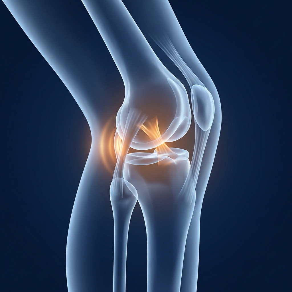
The posterior knee compartment occupies the space between the back of the femur (thigh bone) and the back of the tibia (shin bone). The posterior horns of the medial and lateral menisci sit in this space as shock-absorbing wedges that distribute load and protect cartilage.
The posterior horn of the medial meniscus (PHMM) is the most vulnerable structure in this region. It represents the most common site of meniscal pathology, and deep knee flexion places significant stress on this structure specifically.
The impingement mechanism works as follows: as the knee folds past 120 degrees, the posterior femoral condyle rolls backward and downward, compressing the posterior horn of the medial meniscus between the femur and tibia. When this tissue is torn, degenerated, or mechanically obstructed, the result is sharp, deep posterior pain.
A critical biological fact explains why rest alone rarely resolves this problem. Vascular penetration reaches less than one-third of the adult meniscus. The inner “white zone” where most posterior horn tears occur has severely limited natural healing capacity due to this lack of blood supply.
Meniscal tears have an incidence of 61 per 100,000 in the general U.S. population. Medial meniscal tears occur more commonly than lateral tears, and the posterior horn is the most frequently injured site.
The Three Pathological Culprits Behind Folding Pain
Understanding the three primary structural causes of terminal-flexion pain provides the diagnostic core for addressing this condition effectively.
Posterior Horn Meniscal Tears: The Most Common Starting Point
Posterior horn tears exist on a spectrum including radial tears, peripheral tears, root tears, and the most common subtype: horizontal tears. Medial meniscus posterior horn horizontal tears (MMPHHT) account for 12% to 40% of all meniscus tears and typically occur in individuals over 40 with underlying degenerative changes.
Horizontal tears are particularly associated with deep-flexion pain because the tear plane runs parallel to the tibial plateau, creating a flap that gets pinched during terminal flexion.
The symptom profile includes pain specifically at the back and inner side of the knee during deep squatting, kneeling, or sitting cross-legged. This pain is often accompanied by a sense of catching, clicking, or a dull ache that lingers after the knee is unloaded.
Diagnosis presents challenges. Standard McMurray test sensitivity reaches approximately 70% with specificity around 71%. A “normal” physical exam does not rule out posterior horn pathology, making MRI with a skilled musculoskeletal radiologist the gold standard for confirmation.
Cam Osteophyte Formation: When Bone Becomes the Problem
A cam osteophyte is a bony spur that develops gradually on the posterior part of the medial femoral condyle as a response to chronic impingement stress. This represents the body’s attempt to stabilize a damaged joint, but it instead creates a new mechanical problem.
In a knee with cam impingement of the posterior femoral condyle, the posterior horn of the medial meniscus is pinched and damaged between the medial femoral condyle and the medial tibial plateau during deep flexion. No amount of stretching or strengthening can fully eliminate this compressive force.
Cam osteophyte formation represents the transition from soft-tissue pathology (meniscal tear) to bony-structural pathology. This meaningful escalation changes both the pain character and the treatment calculus.
Research confirms the connection: posterior tibial osteophytes have an odds ratio of 4.525 as an independent contributing factor to medial meniscus posterior horn horizontal tears, demonstrating that bone and soft tissue pathology are deeply interconnected in this syndrome.
Pincer Deformity: The Advanced Structural Stage
In progressive stages of posteromedial impingement, a corresponding bony spur (pincer lesion) develops on the posterior tibia, creating a bony clamp that traps the posterior horn of the meniscus during deep flexion.
At this stage, the posterior horn is mechanically trapped between two bony prominences on every deep flexion movement. The pain becomes more consistent, more severe, and often accompanied by a palpable or audible clunk.
The cam-pincer combination dramatically accelerates cartilage destruction in the posterior compartment. Treatments applied to the meniscus alone will fail without correcting the underlying bony impingement. Resection of the cam lesion may delay progression of knee osteoarthritis, but only if addressed before irreversible cartilage loss has occurred.
The Pathology Cascade: How Folding Pain Becomes Arthritis
The progression follows a clear, sequential framework: posterior horn tear leads to cam osteophyte formation, then pincer deformity, followed by accelerated cartilage destruction, and ultimately compartmental osteoarthritis.
At each stage, the biological mechanism compounds. The torn meniscus loses its ability to distribute load evenly. The exposed cartilage experiences concentrated stress. The body responds with bony remodeling through osteophytes. The osteophytes create new impingement. The cycle accelerates.
The posterior portions of the medial and lateral tibial cartilage are the most vulnerable to osteoarthritis development as a result of deep squatting, precisely the same anatomical zone where posterior horn tears originate.
Over 365 million people globally suffer from knee osteoarthritis. Prevalence has increased by 113% since 1990, and projected figures suggest nearly 1 billion people could be affected by 2050. Knee OA ranks as the third most rapidly rising condition associated with disability, with direct medical costs of $65 billion annually in the United States.
The window for intervention that preserves the joint is real and finite. Patients who dismiss deep-flexion-only pain as “minor” because it does not affect their walking are often the ones who present years later with advanced compartmental osteoarthritis. The absence of pain at rest or during normal gait does not mean the joint is healthy; it means the pathology has not yet progressed to the point of constant load-bearing symptoms.
Three Clinically Validated Self-Diagnostic Tests for Posterior Horn Involvement
These tests replicate what orthopedic specialists use to provoke posterior horn symptoms in clinical settings. Patients can use them to confirm whether their folding pain fits the posterior compartment pattern before seeking formal evaluation.
Safety disclaimer: These tests are for informational purposes and should not replace professional medical evaluation. If any test provokes significant pain, stop immediately.
Ege’s Test (Weight-Bearing Deep Squat Test)
Stand with feet shoulder-width apart and toes pointed outward for medial meniscus testing. Slowly lower into a full squat, holding for a few seconds at the deepest point, then rise.
A positive result includes pain, clicking, or a sense of catching at the posteromedial knee during the deepest phase of the squat, specifically at flexion angles greater than 90 degrees.
Anteriorly-located meniscal tears produce symptoms earlier in knee flexion, whereas tears located on the posterior horn produce symptoms in deeper knee flexion. Ege’s test scored better than McMurray’s for accuracy, sensitivity, and specificity for medial meniscus tears, achieving 81% specificity for medial meniscus tears.
Childress Sign (Duck-Walk Test)
From a full squat position with knees fully bent and buttocks near heels, attempt to walk forward several steps while remaining in the deep squat.
This maneuver provokes compressive force specifically on the posterior horn of the meniscus by maintaining terminal flexion under dynamic load. Pain, a clunk, or a catching sensation at the posteromedial knee during the duck-walk maneuver is highly suggestive of a posterior horn tear.
Many patients with posterior horn tears cannot complete this test at all. The inability to sustain deep flexion under load is itself a clinically meaningful finding.
Payr Sign (Cross-Legged Compression Test)
Sit on the floor or a firm surface in a cross-legged position similar to a meditation posture. Apply gentle downward pressure to the knee that is on top, pushing it toward the floor.
This position places the medial compartment under rotational and compressive stress at a moderate-to-deep flexion angle, specifically loading the posterior horn of the medial meniscus.
Pain at the posteromedial joint line during this maneuver, particularly if it reproduces the patient’s familiar folding pain, represents a positive Payr sign. This test is particularly useful because it replicates the pain pattern of patients who experience symptoms during floor-sitting, yoga, or cross-legged postures.
If two or more of these tests reproduce familiar deep-flexion pain, the probability of posterior horn meniscal involvement is high and warrants formal imaging evaluation.
Why Conservative Care Often Falls Short for Posterior Horn Pathology
Rest, NSAIDs, physical therapy, and activity modification are appropriate first-line interventions and should be attempted. However, they frequently fail for posterior horn tears due to biological and mechanical realities.
The inner zone of the meniscus where most posterior horn tears occur has less than one-third vascular penetration in adults. This tissue has severely limited capacity for self-repair regardless of how much rest is provided.
If a cam osteophyte or pincer lesion is already present, no amount of strengthening or stretching eliminates the bony impingement that occurs at terminal flexion. The structural problem requires a structural solution.
Patients who complete physical therapy and improve general knee strength but do not resolve specific pain during deep flexion encounter a predictable outcome: they are treating a structural problem with a functional intervention. For a broader overview of what is available beyond physical therapy, non-surgical knee treatment options can help patients understand the full spectrum of approaches before committing to a path.
The Science of Regenerative Intervention for Posterior Horn Pathology
For patients at a clinical inflection point after conservative care has failed, understanding the evidence-based rationale for regenerative treatment options becomes essential.
Why the Avascular Posterior Horn Makes Biologics Particularly Relevant
PRP and BMAC deliver concentrated growth factors, platelets, and regenerative signals directly to tissue that cannot access these healing agents through normal blood supply. This essentially bypasses the vascular limitation that makes posterior horn tears so resistant to natural healing.
PRP stimulates cellular proliferation, collagen synthesis, and angiogenesis in the meniscus, the three biological processes most needed in the avascular posterior horn.
A March 2024 systematic review of 10 studies and 686 patients found PRP for degenerative meniscal pathology demonstrated improved pain and knee function within 3 months lasting at least one year, with 30 to 70% of patients radiographically stable and 40 to 60% showing interval healing on MRI.
A meta-analysis of 9 studies with 1,164 participants found that PRP-augmented meniscus repair had a significantly lower failure rate and better postoperative pain control versus non-PRP repair.
A 2025 comprehensive review of 40 high-quality studies found that leukocyte-poor PRP demonstrates superior pain relief and functional improvement compared to hyaluronic acid and corticosteroids, especially in patients with mild to moderate knee OA.
BMAC and Stem Cell Therapy: Addressing the Structural Cascade
BMAC (Bone Marrow Aspiration Concentrate) contains mesenchymal stem cells with the capacity to differentiate into cartilage and fibrocartilage tissue. This becomes relevant for patients who have progressed beyond pure meniscal pathology into early cartilage involvement. Patients interested in understanding how knee cartilage regeneration treatment works as part of this process will find that BMAC plays a central role in addressing cartilage loss alongside meniscal pathology.
The 2026 research landscape continues to validate regenerative approaches. Stanford Medicine scientists discovered that blocking the enzyme 15-PGDH reverses cartilage loss in aging joints and prevents arthritis after knee injuries. Human cartilage samples from knee replacement surgeries also began regenerating when exposed to the treatment.
ARPA-H’s NITRO program is fast-tracking three regenerative approaches for OA in 2026: targeted bone regeneration, targeted cartilage regeneration, and living knee implants made from human tissue. This positions regenerative medicine as the trajectory of orthopedic care rather than an experimental fringe.
As of 2026, the FDA has not approved stem cell, PRP, or exosome products specifically for orthopedic conditions, though same-day autologous procedures remain within FDA regulatory frameworks. Patients seeking clarity on this topic can review the current FDA-approved stem cell therapy orthopedic landscape to understand what regulatory status means for their treatment decisions. This transparency is important for informed patient decision-making.
The Importance of Precision-Guided Delivery for Posterior Compartment Injections
The posterior compartment of the knee is anatomically complex and difficult to access. Biologics delivered to the general joint space may not reach the specific site of posterior horn pathology in sufficient concentration.
Ultrasound-guided knee injection and X-ray guidance for injection procedures ensures accurate delivery of therapeutic agents to the targeted treatment area. This represents a clinical necessity for posterior compartment interventions, not a luxury.
Patients evaluating regenerative providers should specifically ask about imaging-guided injection protocols for posterior compartment treatment.
Who Is a Candidate for Regenerative Intervention?
The ideal regenerative candidate for posterior horn pathology includes patients with confirmed or suspected posterior horn meniscal tears, early cam osteophyte formation, or early-to-moderate compartmental OA who have completed a course of conservative care without adequate resolution of deep-flexion symptoms.
Clinical indicators suggesting regenerative candidacy include persistent posteromedial pain specifically at terminal flexion beyond 120 degrees, positive Ege’s, Childress, or Payr sign, MRI findings consistent with posterior horn pathology, and no or minimal joint space narrowing on weight-bearing X-ray.
Patients with complete meniscal root tears causing joint subluxation, large locked bucket-handle tears, or advanced compartmental OA with severe joint space narrowing may need surgical evaluation first. Regenerative medicine is not a substitute for necessary surgical intervention.
Over 600,000 knee replacements are performed annually in the United States, and studies suggest up to 80% of patients told they need total knee replacement may not actually require surgery. Exploring alternatives to knee replacement surgery early in the posterior horn pathology cascade represents one of the most impactful ways to avoid becoming part of that statistic.
Conclusion: Folding Pain Is a Signal Worth Taking Seriously
Deep-flexion knee pain, the pain that appears specifically when the knee folds past 120 degrees, is not a minor or ambiguous symptom. It is a clinically specific signal pointing to posterior horn meniscal impingement, cam osteophyte formation, or the early stages of a progressive bony-structural cascade toward accelerated osteoarthritis.
The Ege’s test, Childress duck-walk, and Payr sign give patients a concrete way to confirm whether their pain fits the posterior compartment pattern. Two or more positive tests warrant formal evaluation.
The progression from posterior horn tear to cam osteophyte to pincer deformity to cartilage destruction is not inevitable, but it is predictable. The window for intervention that preserves the joint is real.
For patients who have already tried rest, NSAIDs, and physical therapy without resolution, the biological rationale for PRP, BMAC, and precision-guided biologic delivery is well-supported by current clinical evidence. The 2026 research landscape confirms that regenerative medicine represents the direction orthopedic care is moving.
The specificity of this symptom is an advantage, not a mystery. It points directly to the anatomy that needs attention, and that precision makes targeted intervention possible.
Take the Next Step: Evaluate Posterior Horn Pain With Unicorn Bioscience
For individuals experiencing knee pain specifically during deep flexion (squatting, kneeling, sitting cross-legged, or folding the knee fully) where conservative care has not resolved symptoms, a specialized regenerative medicine evaluation can determine whether the symptom profile fits the posterior horn impingement framework.
Unicorn Bioscience specializes in precision-guided regenerative interventions for knee pathology, including PRP, BMAC, and stem cell therapy delivered under ultrasound and X-ray guidance to ensure accurate posterior compartment targeting.
With eight locations across Texas, Florida, and New York, including Austin, Dallas, Houston, San Antonio, Fort Worth, El Paso, Boca Raton, and Manhattan, plus virtual consultation options available, a specialized evaluation is within reach regardless of location.
Qualified candidates can receive injection treatments on the same day as their consultation, minimizing the time between evaluation and intervention.
Contact Unicorn Bioscience at (737) 347-0446 or visit unicornbioscience.com to schedule a virtual or in-person consultation and find out whether deep-flexion knee pain makes regenerative intervention an appropriate option.
More than 90% of Unicorn Bioscience stem cell patients have not gone on to knee replacement surgery, a statistic reflecting what is possible when posterior compartment pathology is identified and treated before the cascade advances.
Schedule Your Consultation Today!
