
Peroneal Tendonitis Regenerative Treatment: The Avascular Zone Framework That Explains Why This Tendon Heals Differently
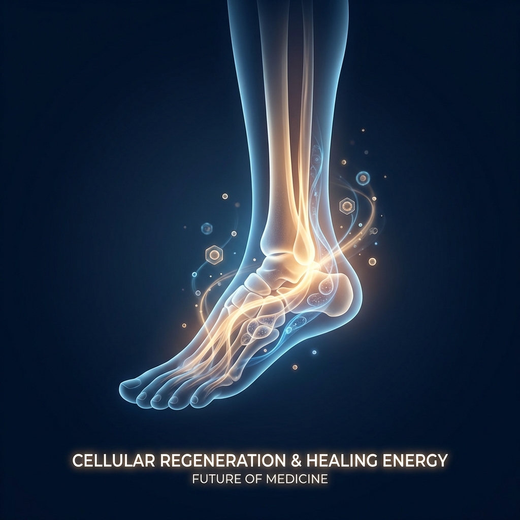
Peroneal Tendonitis Regenerative Treatment: The Avascular Zone Framework That Explains Why This Tendon Heals Differently
Introduction: Why Peroneal Tendonitis Is Not Just Another Ankle Problem
Approximately 60% of peroneal tendonitis patients are initially misdiagnosed, often mistaken for lateral ankle sprains or other lateral ankle pathologies. This striking statistic reveals a fundamental problem in how outer ankle pain is evaluated and treated—and explains why so many patients spend months pursuing ineffective interventions.
The peroneal tendons—peroneus longus and peroneus brevis—run parallel along the outer ankle, wrapping behind the lateral malleolus before diverging toward their respective insertion points. These tendons serve as critical stabilizers of the ankle and enable the foot to evert, or turn outward. When they become inflamed or damaged, the result is persistent lateral ankle pain that can derail athletic careers and compromise daily function.
The central thesis of this article is straightforward: the peroneal tendons contain three anatomically distinct avascular zones that create a biological healing ceiling conventional conservative care cannot overcome. Understanding this framework changes how patients and clinicians should approach treatment.
This article establishes a clear pathway—accurate diagnosis first, understanding why these tendons heal differently second, and how regenerative therapies including PRP, BMAC, and exosomes are uniquely suited to address the avascular zone problem. For active individuals, athletes, runners, dancers, and anyone who has struggled with persistent outer ankle pain despite months of standard treatment, peroneal tendonitis regenerative treatment represents a biologically rational alternative worth understanding.
Understanding Peroneal Tendonitis: Anatomy, Risk Factors, and Who Gets It
The peroneus longus and peroneus brevis run parallel along the outer ankle, wrapping behind the lateral malleolus in a shared fibrous sheath before diverging. The peroneus brevis inserts at the base of the fifth metatarsal, while the peroneus longus continues across the plantar foot to insert at the base of the first metatarsal and medial cuneiform.
The primary causes of peroneal tendonitis include overuse, repetitive ankle movements, sudden inversion sprains, and cumulative microtrauma. The populations most at risk include runners, dancers, soccer and basketball players, and anyone performing frequent cutting or pivoting movements.
Key biomechanical factors that contribute to peroneal tendon pathology include:
- High-arched (cavus) foot that places increased stress on the lateral ankle
- Tight calf muscles that alter ankle mechanics
- Chronic lateral ankle instability from prior sprains
- Improper footwear lacking adequate support
- History of ankle sprains that may have caused unrecognized peroneal damage
A 2025 MRI study of 1,160 ankles identified an important anatomical variant: the peroneus quartus muscle, present in 10.6% of ankles, is significantly associated with peroneus brevis tendinitis and tears due to mechanical crowding in the retromalleolar groove. This finding has direct implications for treatment planning.
Peroneal tendonitis is relatively rare—studies of several thousand runners found fewer than 1% developed the condition. However, its impact on function and quality of life is disproportionate to its prevalence.
Clinicians and patients should distinguish between peroneus brevis tendinopathy, which presents with pain at the lateral malleolus and is associated with retromalleolar groove crowding, and peroneus longus tendinopathy, which produces pain along the lateral foot toward the cuboid notch. This distinction, rarely addressed in standard patient education, affects both diagnosis and treatment selection.
The Misdiagnosis Problem: Why Getting the Right Diagnosis Matters Before Any Treatment
The 60% initial misdiagnosis rate for peroneal tendon injuries demands attention. These injuries are commonly misdiagnosed as lateral ankle sprains, sinus tarsi syndrome, or other lateral ankle pathologies. The clinical consequences of misdiagnosis are significant: inappropriate treatment, delayed healing, progression from tendinitis to partial or full-thickness tears, and wasted time and resources on ineffective interventions.
Accurate diagnostic differentiation requires distinguishing between:
- Tendinitis (inflammatory, early-stage)
- Tendinopathy (degenerative, later-stage)
- Partial or full-thickness tears
- Subluxation (tendon slipping out of the groove)
- Peroneal tunnel syndrome (compression neuropathy)
Each of these conditions requires a different treatment approach. MRI remains the gold standard for identifying structural pathology, while dynamic ultrasound is particularly valuable for detecting subluxation and guiding injections.
Biomechanical analysis is equally important. Core stability, lower extremity alignment, and foot structure must be assessed to identify contributing factors that will affect regenerative treatment outcomes. This diagnostic-first principle is essential: regenerative treatment decisions must be grounded in accurate imaging and clinical assessment.
The Avascular Zone Framework: The Biological Reason Peroneal Tendons Heal Differently
Tendons receive limited blood supply compared to muscle tissue, and regions with reduced or absent vascularity are particularly vulnerable to degenerative change and slow healing. The peroneal tendons contain three distinct avascular zones:
- The zone where the peroneus longus and brevis curve around the posterior lateral malleolus
- The zone where the peroneus longus curves around the cuboid notch
- The retromalleolar groove where mechanical compression further restricts blood flow
In these avascular zones, microtrauma accumulates faster than the tendon can repair itself. This leads to the transition from tendinitis (inflammatory) to tendinopathy (degenerative)—a distinction with major treatment implications.
The failure of conventional care becomes understandable through this framework. Rest, NSAIDs, corticosteroid injections, and physical therapy alone cannot deliver the cellular signals and growth factors needed to initiate repair in tissue with inadequate blood supply. Corticosteroids are particularly problematic in avascular zones: while they reduce inflammation short-term, they do not promote tissue repair and may accelerate collagen degradation in already-compromised tissue.
Tendon healing occurs across three stages: inflammation (days 1–7), cell proliferation (weeks 1–6), and tissue remodeling (months 2–12+). Conventional treatments address only the first stage. Regenerative therapies target the second and third stages—precisely where avascular tendons fail to progress on their own.
If the core problem is insufficient biological signaling in avascular tissue, the solution must involve directly delivering those signals.
How Regenerative Medicine Works for Tendon Healing: The Core Mechanisms
Regenerative medicine for tendons operates on a fundamental principle: rather than suppressing the body’s response or waiting for passive healing, these therapies amplify and direct the body’s own repair mechanisms.
The key cell types involved in tendon repair include tenocytes (tendon-specific cells), tendon stem/progenitor cells (TSCs), and mesenchymal stem cells (MSCs). Each contributes to repair through different mechanisms. Understanding the range of cell types used in regenerative protocols helps patients make informed decisions about their treatment options.
Growth factors play essential roles in tendon repair. PRP concentrates several critical factors:
- PDGF (Platelet-Derived Growth Factor): cell recruitment
- TGF-β (Transforming Growth Factor-beta): extracellular matrix synthesis
- VEGF (Vascular Endothelial Growth Factor): angiogenesis
- EGF, IGF-I, FGF, HGF: proliferation and tissue remodeling
Patient selection is therefore a key determinant of outcomes. Imaging-confirmed diagnosis, disease stage, patient age, activity level, and biomechanical factors all influence which regenerative modality is most appropriate.
Regenerative Treatment Options for Peroneal Tendonitis: A Modality-by-Modality Guide
Each regenerative modality targets a specific phase of tendon healing and is appropriate for a specific stage of disease severity.
PRP (Platelet-Rich Plasma): The First-Line Regenerative Option
PRP is prepared from a small blood draw that is centrifuged to concentrate platelets and their associated growth factors to 3–8x baseline levels. For tendon repair, PRP delivers a concentrated bolus of growth factors directly into the avascular zone, bypassing the blood supply limitation.
Clinical evidence supports PRP efficacy. A multicenter retrospective study of 180 patients treated with ultrasound-guided PRP for chronic tendinopathy—including peroneus longus and brevis—found 82% reported moderate to complete resolution of symptoms, with average VAS pain scores dropping from 7 to 1.8. A 2025 meta-analysis of 16 RCTs found PRP significantly reduced chronic tendinopathy pain with a moderate effect size (SMD = 0.47, p < 0.001).
However, the peroneal-specific evidence requires honest acknowledgment. A separate retrospective study of 214 patients found that while 83% showed overall improvement, VAS scores for peroneal and Achilles tendons specifically did not reach statistical significance at 6 weeks—suggesting peroneal tendons may respond more slowly or require a different protocol than patellar or epicondylar tendons.
PRP formulation variables significantly affect outcomes. Leukocyte-rich versus leukocyte-poor preparations, platelet concentration, and activation method all matter—a key reason why PRP results vary across studies and why provider expertise is important. A detailed understanding of the PRP injection procedure step by step can help patients know what to expect during treatment.
Combining PRP with percutaneous needle tenotomy (tendon fenestration) under ultrasound guidance has shown promising results for chronic recalcitrant tendinopathy, with one study reporting 68% functional improvement and 83% patient satisfaction.
Ultrasound guidance is essential for accurate delivery into the peroneal tendon sheath rather than the surrounding soft tissue.
BMAC (Bone Marrow Aspiration Concentrate): The Advanced Option for Recalcitrant Cases
BMAC involves aspirating bone marrow—typically from the iliac crest—concentrating it, and injecting the concentrate into the damaged tendon. This delivers a rich mixture of mesenchymal stem cells, growth factors, and anti-inflammatory cytokines.
MSCs in BMAC possess multilineage differentiation potential, immunomodulatory activity, and paracrine signaling that promotes angiogenesis and tenocyte proliferation, addressing the avascular zone problem at a cellular level.
BMAC is reserved for recalcitrant cases. The emerging clinical protocol for peroneal tendinopathy positions PRP as the first-line intervention, with BMAC (approximately $3,000) reserved for cases that fail PRP—reflecting both the higher cost and the more invasive aspiration procedure.
Adipose-Derived Stem Cells (ADSCs): An Emerging Alternative
ADSCs are mesenchymal stem cells derived from fat tissue, typically via mini-liposuction, offering a potentially more abundant cell source than bone marrow. Proposed applications in foot and ankle include Achilles tendinopathy, plantar fasciitis, and ankle osteoarthritis, with peroneal tendinopathy as a logical extension.
Exosome Therapy: The Next-Generation Cell-Free Approach
Exosomes are extracellular vesicles (30–150 nm) secreted by MSCs and tenocytes that carry proteins, lipids, and RNA cargo to recipient cells, acting as biological messengers.
For tendon repair, exosomes reduce inflammation, improve collagen organization, enhance biomechanical tendon properties, and stimulate tenocyte proliferation—without the immunogenic risks associated with live cell therapies.
A 2025 review identified tenocyte-derived exosomes as potentially more favorable for tendon repair than MSC-derived exosomes. Preclinical studies show strong results, and early clinical trials suggest safety and potential efficacy.
As a cell-free product, exosomes face a simpler regulatory pathway than live cell therapies and eliminate concerns about immune rejection. Patients interested in understanding how exosome therapy compares to stem cell therapy will find this distinction increasingly relevant as the field advances. They represent a forward-looking option that is not yet standard of care but is advancing rapidly.
The Clinical Protocol: How Regenerative Treatment for Peroneal Tendonitis Is Sequenced
Regenerative medicine specialists follow a structured clinical workflow for peroneal tendinopathy:
Step 1 — Accurate diagnosis: MRI or dynamic ultrasound to confirm tendinitis vs. tear vs. subluxation, rule out peroneus quartus crowding, and assess disease stage.
Step 2 — Biomechanical assessment: Evaluate cavus foot deformity, lateral ankle instability, calf flexibility, and lower extremity alignment.
Step 3 — First-line regenerative intervention: Ultrasound-guided PRP injection into the peroneal tendon sheath, potentially combined with percutaneous needle tenotomy for chronic cases.
Step 4 — Structured rehabilitation: Eccentric loading protocols and progressive tendon loading are essential post-PRP. Regenerative therapy without rehabilitation is suboptimal.
Step 5 — Reassessment at 6–12 weeks: Clinical evaluation and repeat imaging to assess response.
Step 6 — BMAC for recalcitrant cases: Reserved for patients with imaging-confirmed persistent tendinopathy who have failed conservative care and PRP.
Personalized treatment planning—accounting for inflammation levels, patient age, injury type, current medications, and health goals—distinguishes specialized regenerative medicine centers from generic injection clinics. Unicorn Bioscience exemplifies this approach through its personalized regenerative medicine protocols, with all injections administered under ultrasound or X-ray guidance for accurate delivery.
What the Evidence Actually Shows: Honest Expectations for Peroneal Tendonitis Regenerative Treatment
PRP has the strongest clinical evidence base for chronic tendinopathy. However, available evidence suggests peroneal tendons may respond more slowly to PRP than patellar or epicondylar tendons, and patients should expect a longer timeline to full benefit.
Success means meaningful reduction in VAS pain scores, improved functional capacity, return to sport or activity, and avoidance of surgery—not necessarily complete structural normalization on imaging.
Most patients require 3–6 months to experience full benefit, and some may require a second injection cycle. Understanding the PRP therapy recovery timeline helps set realistic expectations for the healing process.
Cost, Insurance, and Practical Considerations
As of 2026, most major insurers classify PRP and stem cell injections as experimental or investigational for musculoskeletal conditions including tendinopathies. The FDA has not approved PRP, stem cell, or exosome products specifically for orthopedic conditions, though substantial clinical evidence supports safety and efficacy when administered by qualified providers within FDA regulatory frameworks.
Current cash-pay price ranges: PRP injections typically range from $500–$2,000 per session; BMAC is approximately $3,000.
For patients who have spent months on physical therapy, corticosteroid injections, and imaging without resolution, regenerative treatment represents a potentially cost-effective alternative to the trajectory toward surgery.
Patients should ask providers about imaging guidance, PRP formulation specifics, and post-injection rehabilitation protocols, as these variables significantly affect outcomes.
Who Is — and Is Not — a Good Candidate for Peroneal Tendonitis Regenerative Treatment
Ideal candidates include:
- Imaging-confirmed peroneal tendinopathy without full-thickness tear
- Failed 3–6 months of conservative care
- Motivated to complete post-injection rehabilitation
- No contraindications to the chosen modality
Patients who may not be ideal candidates:
- Full-thickness peroneal tendon tears requiring surgical repair
- Active infection
- Blood disorders
- Patients on anticoagulants (relative contraindication)
- Advanced-stage tendinopathy with extensive non-tenocyte differentiation
Patients with the peroneus quartus variant may require additional consideration, as mechanical crowding may limit regenerative outcomes without addressing the underlying structural issue. Patients considering treatment should review stem cell injection side effects and risks as part of their informed decision-making process.
The Future of Peroneal Tendon Regeneration: What Is Coming
The frontier of tendon regeneration research includes tissue engineering approaches combining stem cells with biomaterial scaffolds and 3D bioprinting—largely preclinical as of 2026 but advancing rapidly.
With 224 global clinical trials currently investigating stem cell therapies for musculoskeletal conditions and a major Phase III trial funded with $140 million announced in January 2026, the field is maturing rapidly.
Conclusion: The Avascular Zone Is the Problem — Regenerative Medicine Is the Solution
Peroneal tendonitis fails to heal with conventional care not because patients are not trying hard enough, but because the three avascular zones in these tendons create a biological healing ceiling that rest, NSAIDs, and corticosteroids cannot overcome.
Accurate imaging-based diagnosis remains the non-negotiable foundation of any effective treatment plan—the 60% misdiagnosis rate means many patients are treating the wrong problem.
The regenerative treatment hierarchy is clear: PRP as the accessible, evidence-supported first-line option; BMAC for recalcitrant cases; exosomes as an emerging cell-free alternative. Each targets the proliferation and remodeling phases that conventional care cannot reach.
For patients who have exhausted conventional options, peroneal tendonitis regenerative treatment represents a biologically rational, minimally invasive pathway toward recovery.
Take the Next Step: Find Out If Regenerative Treatment Is Right for Your Peroneal Tendonitis
Patients struggling with persistent outer ankle pain may benefit from consulting with regenerative medicine specialists. Unicorn Bioscience offers thorough evaluations that assess inflammation levels, injury type, imaging findings, current medications, and personal health goals—not a one-size-fits-all approach.
Qualified candidates can receive PRP treatment on the same day as their consultation, minimizing delays in care. Virtual consultations are available, making the evaluation process accessible regardless of location.
With clinics in Austin, Dallas, El Paso, Fort Worth, Houston, San Antonio, Boca Raton, and Manhattan, Unicorn Bioscience offers accessible care across Texas, Florida, and New York.
Contact Information:
- Phone: (737) 347-0446
- Website: unicornbioscience.com
Unicorn Bioscience offers imaging-guided, personalized regenerative protocols—PRP, BMAC, and emerging cell-free therapies—as scientifically grounded alternatives to surgery for peroneal tendinopathy.
The longer advanced tendinopathy goes untreated, the greater the risk of progression to structural tear. Early intervention with regenerative therapy offers the best opportunity for full recovery.
Schedule Your Consultation Today!
